7KUF

Transcription activation subcomplex with WhiB7 bound to SigmaAr4-RNAP Beta flap tip chimera and DNA


Experimental Data Snapshot

  • Method: X-RAY DIFFRACTION
  • Resolution: 2.60 Å
  • R-Value Free: 0.255 
  • R-Value Work: 0.221 
  • R-Value Observed: 0.223 

wwPDB Validation   3D Report Full Report


This is version 1.2 of the entry. See complete history


Literature

Structural insights into the functional divergence of WhiB-like proteins in Mycobacterium tuberculosis.

Wan, T.Horova, M.Beltran, D.G.Li, S.Wong, H.X.Zhang, L.M.

(2021) Mol Cell 81: 2887

  • DOI: https://doi.org/10.1016/j.molcel.2021.06.002
  • Primary Citation of Related Structures:  
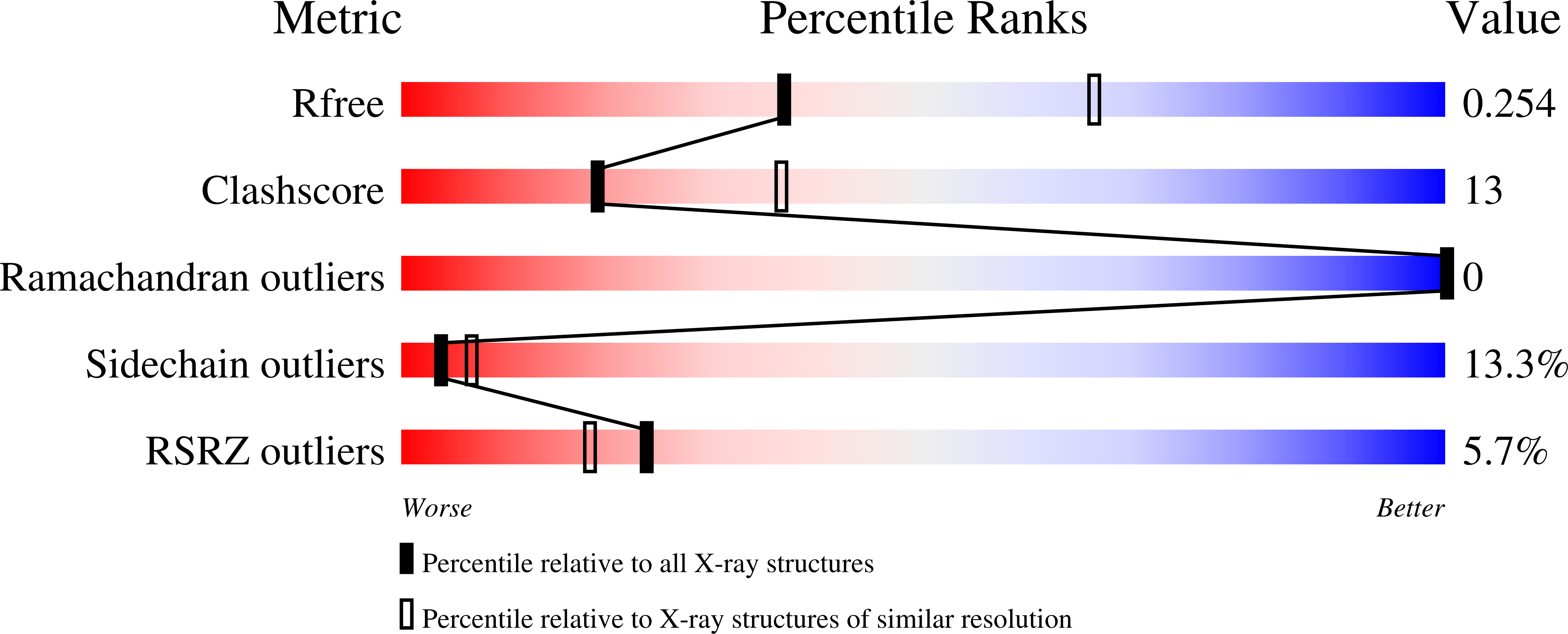
    7KUF, 7KUG

  • PubMed Abstract: 

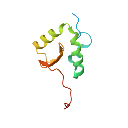
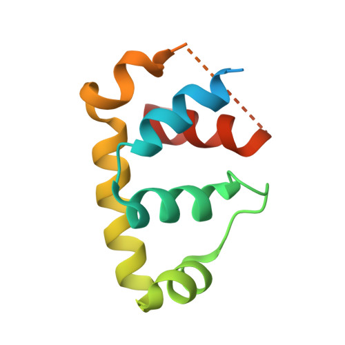
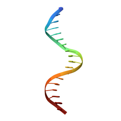
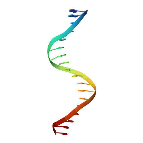
    WhiB7 represents a distinct subclass of transcription factors in the WhiB-Like (Wbl) family, a unique group of iron-sulfur (4Fe-4S] cluster-containing proteins exclusive to the phylum of Actinobacteria. In Mycobacterium tuberculosis (Mtb), WhiB7 interacts with domain 4 of the primary sigma factor (σ A 4 ) in the RNA polymerase holoenzyme and activates genes involved in multiple drug resistance and redox homeostasis. Here, we report crystal structures of the WhiB7:σ A 4 complex alone and bound to its target promoter DNA at 1.55-Å and 2.6-Å resolution, respectively. These structures show how WhiB7 regulates gene expression by interacting with both σ A 4 and the AT-rich sequence upstream of the -35 promoter DNA via its C-terminal DNA-binding motif, the AT-hook. By combining comparative structural analysis of the two high-resolution σ A 4 -bound Wbl structures with molecular and biochemical approaches, we identify the structural basis of the functional divergence between the two distinct subclasses of Wbl proteins in Mtb.


  • Organizational Affiliation

    Department of Biochemistry, University of Nebraska-Lincoln, Lincoln, NE 68588, USA.


Macromolecules

Find similar proteins by:  (by identity cutoff)  |  3D Structure
Entity ID: 1
MoleculeChains Sequence LengthOrganismDetailsImage
Probable transcriptional regulator WhiB792Mycobacterium tuberculosisMutation(s): 0 
Gene Names: whiB7Rv3197ARv3197.1
UniProt
Find proteins for Q6MX01 (Mycobacterium tuberculosis (strain ATCC 25618 / H37Rv))
Explore Q6MX01 
Go to UniProtKB:  Q6MX01
Entity Groups  
Sequence Clusters30% Identity50% Identity70% Identity90% Identity95% Identity100% Identity
UniProt GroupQ6MX01
Sequence Annotations
Expand
  • Reference Sequence
Find similar proteins by:  (by identity cutoff)  |  3D Structure
Entity ID: 2
MoleculeChains Sequence LengthOrganismDetailsImage
RNA polymerase sigma factor, DNA-directed RNA polymerase subunit beta chimera112Mycobacterium tuberculosisMutation(s): 0 
Gene Names: sigAERS007670_01286ERS007681_03833ERS007688_01862
EC: 2.7.7.6
UniProt
Find proteins for P9WGY9 (Mycobacterium tuberculosis (strain ATCC 25618 / H37Rv))
Explore P9WGY9 
Go to UniProtKB:  P9WGY9
Find proteins for P9WGI1 (Mycobacterium tuberculosis (strain ATCC 25618 / H37Rv))
Explore P9WGI1 
Go to UniProtKB:  P9WGI1
Entity Groups  
Sequence Clusters30% Identity50% Identity70% Identity90% Identity95% Identity100% Identity
UniProt GroupsP9WGY9P9WGI1
Sequence Annotations
Expand
  • Reference Sequence

Find similar nucleic acids by:  Sequence   |   3D Structure  

Entity ID: 3
MoleculeChains LengthOrganismImage
DNA (5'-D(*CP*AP*GP*AP*AP*AP*AP*TP*CP*GP*GP*TP*TP*GP*TP*GP*GP*T)-3')18Mycobacterium tuberculosis
Sequence Annotations
Expand
  • Reference Sequence

Find similar nucleic acids by:  Sequence   |   3D Structure  

Entity ID: 4
MoleculeChains LengthOrganismImage
DNA (5'-D(*GP*AP*CP*CP*AP*CP*AP*AP*CP*CP*GP*AP*TP*TP*TP*TP*CP*T)-3')18Mycobacterium tuberculosis
Sequence Annotations
Expand
  • Reference Sequence
Small Molecules
Ligands 1 Unique
IDChains Name / Formula / InChI Key2D Diagram3D Interactions
SF4 (Subject of Investigation/LOI)
Query on SF4

Download Ideal Coordinates CCD File 
E [auth A]IRON/SULFUR CLUSTER
Fe4 S4
LJBDFODJNLIPKO-UHFFFAOYSA-N
Experimental Data & Validation

Experimental Data

  • Method: X-RAY DIFFRACTION
  • Resolution: 2.60 Å
  • R-Value Free: 0.255 
  • R-Value Work: 0.221 
  • R-Value Observed: 0.223 
  • Space Group: P 32 2 1
Unit Cell:
Length ( Å )Angle ( ˚ )
a = 69.254α = 90
b = 69.254β = 90
c = 167.374γ = 120
Software Package:
Software NamePurpose
PHENIXrefinement
HKL-3000data reduction
HKL-3000data scaling
PHASERphasing

Structure Validation

View Full Validation Report


Entry History & Funding Information

Deposition Data


Funding OrganizationLocationGrant Number
National Science Foundation (NSF, United States)United States2562300224001
National Institutes of Health/National Institute of General Medical Sciences (NIH/NIGMS)United StatesR35 GM138157-01
National Institutes of Health/National Institute of General Medical Sciences (NIH/NIGMS)United StatesP20 GM113126
National Institutes of Health/National Institute of General Medical Sciences (NIH/NIGMS)United StatesP30 GM103335

Revision History  (Full details and data files)

  • Version 1.0: 2021-06-30
    Type: Initial release
  • Version 1.1: 2021-10-06
    Changes: Database references
  • Version 1.2: 2023-10-18
    Changes: Data collection, Refinement description