7F8K

Room temperature structure of bacterial copper amine oxidase determined by serial femtosecond crystallography


Experimental Data Snapshot

  • Method: X-RAY DIFFRACTION
  • Resolution: 2.20 Å
  • R-Value Free: 0.179 
  • R-Value Work: 0.154 
  • R-Value Observed: 0.155 

wwPDB Validation   3D Report Full Report


This is version 1.1 of the entry. See complete history


Literature

Microcrystal preparation for serial femtosecond X-ray crystallography of bacterial copper amine oxidase

Murakawa, T.Suzuki, M.Arima, T.Sugahara, M.Tanaka, T.Tanaka, R.Iwata, S.Nango, E.Tono, K.Hayashi, H.Fukui, K.Yano, T.Tanizawa, K.Okajima, T.

(2021) Acta Crystallogr F Struct Biol Commun 77: 356-363

  • DOI: https://doi.org/10.1107/S2053230X21008967
  • Primary Citation of Related Structures:  
    7F8K

  • PubMed Abstract: 

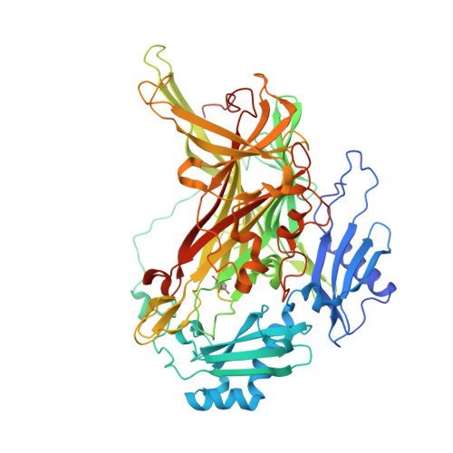
    Recent advances in serial femtosecond X-ray crystallography (SFX) using X-ray free-electron lasers have paved the way for determining radiation-damage-free protein structures under nonfreezing conditions. However, the large-scale preparation of high-quality microcrystals of uniform size is a prerequisite for SFX, and this has been a barrier to its widespread application. Here, a convenient method for preparing high-quality microcrystals of a bacterial quinoprotein enzyme, copper amine oxidase from Arthrobacter globiformis, is reported. The method consists of the mechanical crushing of large crystals (5-15 mm 3 ), seeding the crushed crystals into the enzyme solution and standing for 1 h at an ambient temperature of ∼26°C, leading to the rapid formation of microcrystals with a uniform size of 3-5 µm. The microcrystals diffracted X-rays to a resolution beyond 2.0 Å in SFX measurements at the SPring-8 Angstrom Compact Free Electron Laser facility. The damage-free structure determined at 2.2 Å resolution was essentially identical to that determined previously by cryogenic crystallography using synchrotron X-ray radiation.


  • Organizational Affiliation

    Department of Biochemistry, Faculty of Medicine, Osaka Medical and Pharmaceutical University, 2-7 Daigakumachi, Takatsuki, Osaka 569-8686, Japan.


Macromolecules
Find similar proteins by:  (by identity cutoff)  |  3D Structure
Entity ID: 1
MoleculeChains Sequence LengthOrganismDetailsImage
Phenylethylamine oxidase638Arthrobacter globiformisMutation(s): 0 
EC: 1.4.3.21
UniProt
Find proteins for P46881 (Arthrobacter globiformis)
Explore P46881 
Go to UniProtKB:  P46881
Entity Groups  
Sequence Clusters30% Identity50% Identity70% Identity90% Identity95% Identity100% Identity
UniProt GroupP46881
Sequence Annotations
Expand
  • Reference Sequence
Small Molecules
Ligands 1 Unique
IDChains Name / Formula / InChI Key2D Diagram3D Interactions
CU
Query on CU

Download Ideal Coordinates CCD File 
B [auth A]COPPER (II) ION
Cu
JPVYNHNXODAKFH-UHFFFAOYSA-N
Modified Residues  1 Unique
IDChains TypeFormula2D DiagramParent
TPQ
Query on TPQ
A
L-PEPTIDE LINKINGC9 H9 N O5TYR
Experimental Data & Validation

Experimental Data

  • Method: X-RAY DIFFRACTION
  • Resolution: 2.20 Å
  • R-Value Free: 0.179 
  • R-Value Work: 0.154 
  • R-Value Observed: 0.155 
  • Space Group: C 1 2 1
Unit Cell:
Length ( Å )Angle ( ˚ )
a = 158.75α = 90
b = 64.74β = 112.33
c = 93.55γ = 90
Software Package:
Software NamePurpose
PHENIXrefinement
CrystFELdata reduction
CrystFELdata scaling
PHASESphasing

Structure Validation

View Full Validation Report


Entry History & Funding Information

Deposition Data


Funding OrganizationLocationGrant Number
Japan Society for the Promotion of Science (JSPS)Japan20H05448

Revision History  (Full details and data files)

  • Version 1.0: 2021-09-08
    Type: Initial release
  • Version 1.1: 2021-10-13
    Changes: Database references