7A2D

Structure-function analyses of dual-BON domain protein DolP identifies phospholipid binding as a new mechanism for protein localisation to the cell division site


Experimental Data Snapshot

  • Method: SOLUTION NMR
  • Conformers Calculated: 100 
  • Conformers Submitted: 20 
  • Selection Criteria: structures with the lowest energy 

wwPDB Validation   3D Report Full Report


This is version 1.3 of the entry. See complete history


Literature

Structure of dual BON-domain protein DolP identifies phospholipid binding as a new mechanism for protein localisation.

Bryant, J.A.Morris, F.C.Knowles, T.J.Maderbocus, R.Heinz, E.Boelter, G.Alodaini, D.Colyer, A.Wotherspoon, P.J.Staunton, K.A.Jeeves, M.Browning, D.F.Sevastsyanovich, Y.R.Wells, T.J.Rossiter, A.E.Bavro, V.N.Sridhar, P.Ward, D.G.Chong, Z.S.Goodall, E.C.Icke, C.Teo, A.C.Chng, S.S.Roper, D.I.Lithgow, T.Cunningham, A.F.Banzhaf, M.Overduin, M.Henderson, I.R.

(2020) Elife 9

  • DOI: https://doi.org/10.7554/eLife.62614
  • Primary Citation of Related Structures:  
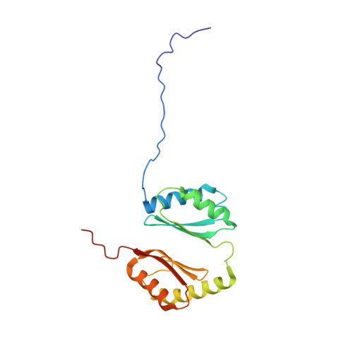
    7A2D

  • PubMed Abstract: 

    The Gram-negative outer-membrane envelops the bacterium and functions as a permeability barrier against antibiotics, detergents, and environmental stresses. Some virulence factors serve to maintain the integrity of the outer membrane, including DolP (formerly YraP) a protein of unresolved structure and function. Here, we reveal DolP is a lipoprotein functionally conserved amongst Gram-negative bacteria and that loss of DolP increases membrane fluidity. We present the NMR solution structure for Escherichia coli DolP, which is composed of two BON domains that form an interconnected opposing pair. The C-terminal BON domain binds anionic phospholipids through an extensive membrane:protein interface. This interaction is essential for DolP function and is required for sub-cellular localisation of the protein to the cell division site, providing evidence of subcellular localisation of these phospholipids within the outer membrane. The structure of DolP provides a new target for developing therapies that disrupt the integrity of the bacterial cell envelope.


  • Organizational Affiliation

    Institute of Microbiology and Infection, University of Birmingham, Edgbaston, United Kingdom.


Macromolecules
Find similar proteins by:  (by identity cutoff)  |  3D Structure
Entity ID: 1
MoleculeChains Sequence LengthOrganismDetailsImage
Uncharacterized protein YraP182Escherichia coli K-12Mutation(s): 0 
Gene Names: yraPb3150JW3119WCM_00535
UniProt
Find proteins for P64596 (Escherichia coli (strain K12))
Explore P64596 
Go to UniProtKB:  P64596
Entity Groups  
Sequence Clusters30% Identity50% Identity70% Identity90% Identity95% Identity100% Identity
UniProt GroupP64596
Sequence Annotations
Expand
  • Reference Sequence
Experimental Data & Validation

Experimental Data

  • Method: SOLUTION NMR
  • Conformers Calculated: 100 
  • Conformers Submitted: 20 
  • Selection Criteria: structures with the lowest energy 

Structure Validation

View Full Validation Report


Entry History & Funding Information

Deposition Data


Funding OrganizationLocationGrant Number
Biotechnology and Biological Sciences Research Council (BBSRC)United KingdomBB/M00810X/1
Biotechnology and Biological Sciences Research Council (BBSRC)United KingdomBB/L00335X/1
Biotechnology and Biological Sciences Research Council (BBSRC)United KingdomBB/P009840/1
Natural Sciences and Engineering Research Council (NSERC, Canada)CanadaRGPIN-2018-04994

Revision History  (Full details and data files)

  • Version 1.0: 2020-12-30
    Type: Initial release
  • Version 1.1: 2021-01-20
    Changes: Database references, Source and taxonomy, Structure summary
  • Version 1.2: 2021-01-27
    Changes: Database references
  • Version 1.3: 2021-02-10
    Changes: Database references