6ZSU

Structure of crocagin biosynthetic protein CgnE


Experimental Data Snapshot

  • Method: X-RAY DIFFRACTION
  • Resolution: 2.00 Å
  • R-Value Free: 0.202 
  • R-Value Work: 0.156 
  • R-Value Observed: 0.159 

wwPDB Validation   3D Report Full Report


This is version 1.2 of the entry. See complete history


Literature

Unusual peptide-binding proteins guide pyrroloindoline alkaloid formation in crocagin biosynthesis.

Adam, S.Zheng, D.Klein, A.Volz, C.Mullen, W.Shirran, S.L.Smith, B.O.Kalinina, O.V.Muller, R.Koehnke, J.

(2023) Nat Chem 15: 560-568

  • DOI: https://doi.org/10.1038/s41557-023-01153-w
  • Primary Citation of Related Structures:  
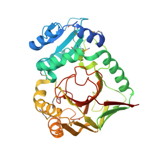
    6ZSU, 6ZSV, 7PD7, 8A2N

  • PubMed Abstract: 

    Ribosomally synthesized and post-translationally modified peptide natural products have provided many highly unusual scaffolds. This includes the intriguing alkaloids crocagins, which possess a tetracyclic core structure and whose biosynthesis has remained enigmatic. Here we use in vitro experiments to demonstrate that three proteins, CgnB, CgnC and CgnE, are sufficient for the production of the hallmark tetracyclic crocagin core from the precursor peptide CgnA. The crystal structures of the homologues CgnB and CgnE reveal them to be the founding members of a peptide-binding protein family and allow us to rationalize their distinct functions. We further show that the hydrolase CgnD liberates the crocagin core scaffold, which is subsequently N-methylated by CgnL. These insights allow us to propose a biosynthetic scheme for crocagins. Bioinformatic analyses based on these data led to the discovery of related biosynthetic pathways that may provide access to a structurally diverse family of peptide-derived pyrroloindoline alkaloids.


  • Organizational Affiliation

    Workgroup Structural Biology of Biosynthetic Enzymes, Helmholtz Institute for Pharmaceutical Research Saarland (HIPS), Helmholtz Centre for Infection Research (HZI), Saarland University, Saarbrücken, Germany.


Macromolecules
Find similar proteins by:  (by identity cutoff)  |  3D Structure
Entity ID: 1
MoleculeChains Sequence LengthOrganismDetailsImage
CgnE
A, B
326Chondromyces crocatusMutation(s): 0 
Gene Names: CMC5_025530
UniProt
Find proteins for A0A0K1ECI7 (Chondromyces crocatus)
Explore A0A0K1ECI7 
Go to UniProtKB:  A0A0K1ECI7
Entity Groups  
Sequence Clusters30% Identity50% Identity70% Identity90% Identity95% Identity100% Identity
UniProt GroupA0A0K1ECI7
Sequence Annotations
Expand
  • Reference Sequence
Small Molecules
Modified Residues  1 Unique
IDChains TypeFormula2D DiagramParent
MSE
Query on MSE
A, B
L-PEPTIDE LINKINGC5 H11 N O2 SeMET
Experimental Data & Validation

Experimental Data

  • Method: X-RAY DIFFRACTION
  • Resolution: 2.00 Å
  • R-Value Free: 0.202 
  • R-Value Work: 0.156 
  • R-Value Observed: 0.159 
  • Space Group: P 21 21 21
Unit Cell:
Length ( Å )Angle ( ˚ )
a = 70.4α = 90
b = 75.34β = 90
c = 112.44γ = 90
Software Package:
Software NamePurpose
PHENIXphasing
PHENIXrefinement
XDSdata reduction
SCALAdata scaling
Cootmodel building

Structure Validation

View Full Validation Report


Entry History & Funding Information

Deposition Data


Funding OrganizationLocationGrant Number
German Research Foundation (DFG)GermanyKO 4116/3-2

Revision History  (Full details and data files)

  • Version 1.0: 2022-07-20
    Type: Initial release
  • Version 1.1: 2023-02-22
    Changes: Database references
  • Version 1.2: 2024-02-28
    Changes: Data collection, Database references