6Y2H

The crystal structure of human chloride intracellular channel protein 5


Experimental Data Snapshot

  • Method: X-RAY DIFFRACTION
  • Resolution: 2.15 Å
  • R-Value Free: 0.244 
  • R-Value Work: 0.201 
  • R-Value Observed: 0.203 

wwPDB Validation   3D Report Full Report


This is version 1.5 of the entry. See complete history


Literature

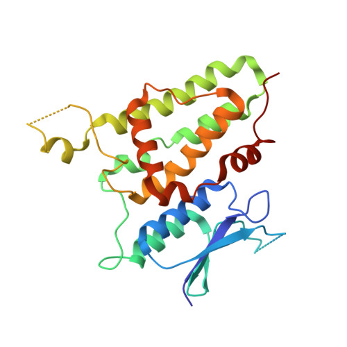
Conserved cysteine dioxidation enhances membrane interaction of human Cl - intracellular channel 5.

Ferofontov, A.Vankova, P.Man, P.Giladi, M.Haitin, Y.

(2020) FASEB J 34: 9925-9940

  • DOI: https://doi.org/10.1096/fj.202000399R
  • Primary Citation of Related Structures:  
    6Y2H

  • PubMed Abstract: 

    The human chloride intracellular channel (hCLIC) family is thought to transition between globular and membrane-associated forms by exposure of a hydrophobic surface. However, the molecular identity of this surface, and the triggering events leading to its exposure, remain elusive. Here, by combining biochemical and structural approaches, together with mass spectrometry (MS) analyses, we show that hCLIC5 is inherently flexible. X-ray crystallography revealed the existence of a globular conformation, while small-angle X-ray scattering showed additional elongated forms consisting of exposure of the conserved hydrophobic inter-domain interface to the bulk phase. Tryptophan fluorescence measurements demonstrated that the transition to the membrane-associated form is enhanced by the presence of oxidative environment and lipids. Using MS, we identified a dose-dependent oxidation of a highly conserved cysteine residue, known to play a key role in the structurally related omega-class of glutathione-S-transferases. Hydrogen/deuterium exchange MS analysis revealed that oxidation of this cysteine facilitates the exposure of the conserved hydrophobic inter-domain interface. Together, our results pinpoint an oxidation of a specific cysteine residue as a triggering mechanism initializing the molecular commitment for membrane interaction in the CLIC family.


  • Organizational Affiliation

    Department of Physiology and Pharmacology, Sackler School of Medicine, Tel-Aviv University, Tel-Aviv, Israel.


Macromolecules
Find similar proteins by:  (by identity cutoff)  |  3D Structure
Entity ID: 1
MoleculeChains Sequence LengthOrganismDetailsImage
Chloride intracellular channel protein 5
A, B, C, D
240Homo sapiensMutation(s): 0 
Gene Names: CLIC5
UniProt & NIH Common Fund Data Resources
Find proteins for Q9NZA1 (Homo sapiens)
Explore Q9NZA1 
Go to UniProtKB:  Q9NZA1
PHAROS:  Q9NZA1
GTEx:  ENSG00000112782 
Entity Groups  
Sequence Clusters30% Identity50% Identity70% Identity90% Identity95% Identity100% Identity
UniProt GroupQ9NZA1
Sequence Annotations
Expand
  • Reference Sequence
Small Molecules
Ligands 1 Unique
IDChains Name / Formula / InChI Key2D Diagram3D Interactions
SCN
Query on SCN

Download Ideal Coordinates CCD File 
E [auth A]
F [auth A]
G [auth A]
H [auth B]
I [auth B]
E [auth A],
F [auth A],
G [auth A],
H [auth B],
I [auth B],
J [auth B],
K [auth B],
L [auth B],
M [auth B],
N [auth C],
O [auth C],
P [auth C],
Q [auth D]
THIOCYANATE ION
C N S
ZMZDMBWJUHKJPS-UHFFFAOYSA-M
Experimental Data & Validation

Experimental Data

  • Method: X-RAY DIFFRACTION
  • Resolution: 2.15 Å
  • R-Value Free: 0.244 
  • R-Value Work: 0.201 
  • R-Value Observed: 0.203 
  • Space Group: C 1 2 1
Unit Cell:
Length ( Å )Angle ( ˚ )
a = 130.99α = 90
b = 93.96β = 124.13
c = 109.58γ = 90
Software Package:
Software NamePurpose
XSCALEdata scaling
PHENIXrefinement
PDB_EXTRACTdata extraction
XSCALEdata reduction
PHENIXphasing

Structure Validation

View Full Validation Report


Entry History & Funding Information

Deposition Data


Funding OrganizationLocationGrant Number
Israel Science FoundationIsrael1721/16
Israel Science FoundationIsrael1775/12
German-Israeli Foundation for Research and DevelopmentIsraelI-2425-418.13/2016
Other privateIsraelICRF 01214

Revision History  (Full details and data files)

  • Version 1.0: 2020-05-20
    Type: Initial release
  • Version 1.1: 2020-08-26
    Changes: Database references
  • Version 1.2: 2020-09-02
    Changes: Database references
  • Version 1.3: 2020-09-30
    Changes: Database references
  • Version 1.4: 2020-12-02
    Changes: Database references
  • Version 1.5: 2024-01-24
    Changes: Data collection, Database references, Refinement description