6WUK

Crystal structure of mouse DXO in complex with CoA


Experimental Data Snapshot

  • Method: X-RAY DIFFRACTION
  • Resolution: 1.60 Å
  • R-Value Free: 0.214 
  • R-Value Work: 0.185 
  • R-Value Observed: 0.186 

wwPDB Validation   3D Report Full Report


Ligand Structure Quality Assessment 


This is version 1.2 of the entry. See complete history


Literature

DXO/Rai1 enzymes remove 5'-end FAD and dephospho-CoA caps on RNAs.

Doamekpor, S.K.Grudzien-Nogalska, E.Mlynarska-Cieslak, A.Kowalska, J.Kiledjian, M.Tong, L.

(2020) Nucleic Acids Res 48: 6136-6148

  • DOI: https://doi.org/10.1093/nar/gkaa297
  • Primary Citation of Related Structures:  
    6WUF, 6WUG, 6WUI, 6WUK

  • PubMed Abstract: 

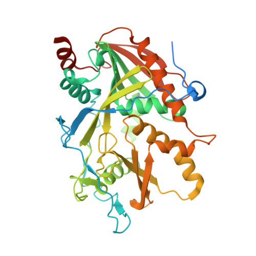
    In eukaryotes, the DXO/Rai1 enzymes can eliminate most of the incomplete and non-canonical NAD caps through their decapping, deNADding and pyrophosphohydrolase activities. Here, we report that these enzymes can also remove FAD and dephospho-CoA (dpCoA) non-canonical caps from RNA, and we have named these activities deFADding and deCoAping. The crystal structures of mammalian DXO with 3'-FADP or CoA and fission yeast Rai1 with 3'-FADP provide elegant insight to these activities. FAD and CoA are accommodated in the DXO/Rai1 active site by adopting folded conformations. The flavin of FAD and the pantetheine group of CoA contact the same region at the bottom of the active site tunnel, which undergoes conformational changes to accommodate the different cap moieties. We have developed FAD-capQ to detect and quantify FAD-capped RNAs and determined that FAD caps are present on short RNAs (with less than ∼200 nucleotides) in human cells and that these RNAs are stabilized in the absence of DXO.


  • Organizational Affiliation

    Department of Biological Sciences, Columbia University, New York, NY 10027, USA.


Macromolecules
Find similar proteins by:  (by identity cutoff)  |  3D Structure
Entity ID: 1
MoleculeChains Sequence LengthOrganismDetailsImage
Decapping and exoribonuclease protein417Mus musculusMutation(s): 3 
Gene Names: DxoDom3zNg6
EC: 3.1.13 (PDB Primary Data), 3.6.1 (PDB Primary Data)
UniProt
Find proteins for O70348 (Mus musculus)
Explore O70348 
Go to UniProtKB:  O70348
Entity Groups  
Sequence Clusters30% Identity50% Identity70% Identity90% Identity95% Identity100% Identity
UniProt GroupO70348
Sequence Annotations
Expand
  • Reference Sequence
Small Molecules
Ligands 1 Unique
IDChains Name / Formula / InChI Key2D Diagram3D Interactions
COA (Subject of Investigation/LOI)
Query on COA

Download Ideal Coordinates CCD File 
B [auth A]COENZYME A
C21 H36 N7 O16 P3 S
RGJOEKWQDUBAIZ-IBOSZNHHSA-N
Experimental Data & Validation

Experimental Data

  • Method: X-RAY DIFFRACTION
  • Resolution: 1.60 Å
  • R-Value Free: 0.214 
  • R-Value Work: 0.185 
  • R-Value Observed: 0.186 
  • Space Group: P 1 21 1
Unit Cell:
Length ( Å )Angle ( ˚ )
a = 49.862α = 90
b = 87.665β = 112.24
c = 54.023γ = 90
Software Package:
Software NamePurpose
XDSdata scaling
PHENIXrefinement
PDB_EXTRACTdata extraction
XDSdata reduction
PHENIXphasing

Structure Validation

View Full Validation Report


Ligand Structure Quality Assessment 


Entry History & Funding Information

Deposition Data


Funding OrganizationLocationGrant Number
National Institutes of Health/National Library of Medicine (NIH/NLM)United StatesR35GM118093

Revision History  (Full details and data files)

  • Version 1.0: 2020-05-20
    Type: Initial release
  • Version 1.1: 2020-06-24
    Changes: Database references
  • Version 1.2: 2023-10-18
    Changes: Data collection, Database references, Refinement description