6VW7

Formate Dehydrogenase FdsABG subcomplex FdsBG from C. necator - NADH bound


Experimental Data Snapshot

  • Method: X-RAY DIFFRACTION
  • Resolution: 2.00 Å
  • R-Value Free: 0.199 
  • R-Value Work: 0.159 
  • R-Value Observed: 0.160 

wwPDB Validation   3D Report Full Report


Ligand Structure Quality Assessment 


This is version 1.3 of the entry. See complete history


Literature

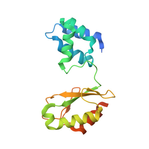
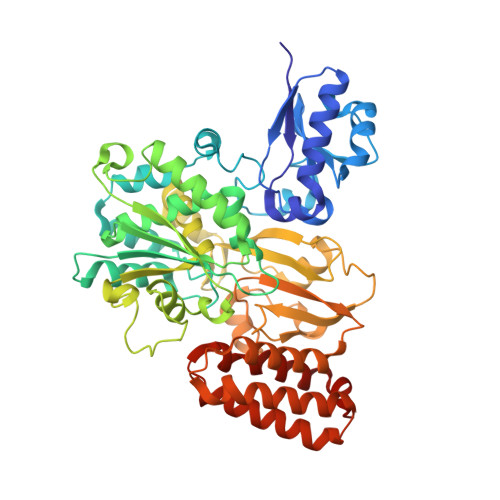
Crystallographic and kinetic analyses of the FdsBG subcomplex of the cytosolic formate dehydrogenase FdsABG fromCupriavidus necator.

Young, T.Niks, D.Hakopian, S.Tam, T.K.Yu, X.Hille, R.Blaha, G.M.

(2020) J Biol Chem 295: 6570-6585

  • DOI: https://doi.org/10.1074/jbc.RA120.013264
  • Primary Citation of Related Structures:  
    6VW7, 6VW8

  • PubMed Abstract: 

    Formate oxidation to carbon dioxide is a key reaction in one-carbon compound metabolism, and its reverse reaction represents the first step in carbon assimilation in the acetogenic and methanogenic branches of many anaerobic organisms. The molybdenum-containing dehydrogenase FdsABG is a soluble NAD + -dependent formate dehydrogenase and a member of the NADH dehydrogenase superfamily. Here, we present the first structure of the FdsBG subcomplex of the cytosolic FdsABG formate dehydrogenase from the hydrogen-oxidizing bacterium Cupriavidus necator H16 both with and without bound NADH. The structures revealed that the two iron-sulfur clusters, Fe 4 S 4 in FdsB and Fe 2 S 2 in FdsG, are closer to the FMN than they are in other NADH dehydrogenases. Rapid kinetic studies and EPR measurements of rapid freeze-quenched samples of the NADH reduction of FdsBG identified a neutral flavin semiquinone, FMNH , not previously observed to participate in NADH-mediated reduction of the FdsABG holoenzyme. We found that this semiquinone forms through the transfer of one electron from the fully reduced FMNH - , initially formed via NADH-mediated reduction, to the Fe 2 S 2 cluster. This Fe 2 S 2 cluster is not part of the on-path chain of iron-sulfur clusters connecting the FMN of FdsB with the active-site molybdenum center of FdsA. According to the NADH-bound structure, the nicotinamide ring stacks onto the re -face of the FMN. However, NADH binding significantly reduced the electron density for the isoalloxazine ring of FMN and induced a conformational change in residues of the FMN-binding pocket that display peptide-bond flipping upon NAD + binding in proper NADH dehydrogenases.


  • Organizational Affiliation

    Department of Biochemistry, University of California, Riverside, California 92521.


Macromolecules
Find similar proteins by:  (by identity cutoff)  |  3D Structure
Entity ID: 1
MoleculeChains Sequence LengthOrganismDetailsImage
NAD-dependent formate dehydrogenase gamma subunit
A, C
176Cupriavidus necatorMutation(s): 0 
Gene Names: fdsGH16_A0640
EC: 1.2.1.2
UniProt
Find proteins for Q0KDY3 (Cupriavidus necator (strain ATCC 17699 / DSM 428 / KCTC 22496 / NCIMB 10442 / H16 / Stanier 337))
Explore Q0KDY3 
Go to UniProtKB:  Q0KDY3
Entity Groups  
Sequence Clusters30% Identity50% Identity70% Identity90% Identity95% Identity100% Identity
UniProt GroupQ0KDY3
Sequence Annotations
Expand
  • Reference Sequence
Find similar proteins by:  (by identity cutoff)  |  3D Structure
Entity ID: 2
MoleculeChains Sequence LengthOrganismDetailsImage
NAD-dependent formate dehydrogenase beta subunit
B, D
520Cupriavidus necatorMutation(s): 0 
Gene Names: fdsBH16_A0641
EC: 1.2.1.2
UniProt
Find proteins for Q0KDY2 (Cupriavidus necator (strain ATCC 17699 / DSM 428 / KCTC 22496 / NCIMB 10442 / H16 / Stanier 337))
Explore Q0KDY2 
Go to UniProtKB:  Q0KDY2
Entity Groups  
Sequence Clusters30% Identity50% Identity70% Identity90% Identity95% Identity100% Identity
UniProt GroupQ0KDY2
Sequence Annotations
Expand
  • Reference Sequence
Small Molecules
Ligands 4 Unique
IDChains Name / Formula / InChI Key2D Diagram3D Interactions
NAI (Subject of Investigation/LOI)
Query on NAI

Download Ideal Coordinates CCD File 
H [auth B],
L [auth D]
1,4-DIHYDRONICOTINAMIDE ADENINE DINUCLEOTIDE
C21 H29 N7 O14 P2
BOPGDPNILDQYTO-NNYOXOHSSA-N
FMN (Subject of Investigation/LOI)
Query on FMN

Download Ideal Coordinates CCD File 
F [auth B],
J [auth D]
FLAVIN MONONUCLEOTIDE
C17 H21 N4 O9 P
FVTCRASFADXXNN-SCRDCRAPSA-N
SF4
Query on SF4

Download Ideal Coordinates CCD File 
G [auth B],
K [auth D]
IRON/SULFUR CLUSTER
Fe4 S4
LJBDFODJNLIPKO-UHFFFAOYSA-N
FES
Query on FES

Download Ideal Coordinates CCD File 
E [auth A],
I [auth C]
FE2/S2 (INORGANIC) CLUSTER
Fe2 S2
NIXDOXVAJZFRNF-UHFFFAOYSA-N
Binding Affinity Annotations 
IDSourceBinding Affinity
NAI Binding MOAD:  6VW7 Kd: 1.90e+5 (nM) from 1 assay(s)
Experimental Data & Validation

Experimental Data

  • Method: X-RAY DIFFRACTION
  • Resolution: 2.00 Å
  • R-Value Free: 0.199 
  • R-Value Work: 0.159 
  • R-Value Observed: 0.160 
  • Space Group: C 1 2 1
Unit Cell:
Length ( Å )Angle ( ˚ )
a = 132.656α = 90
b = 127.05β = 123.5
c = 99.84γ = 90
Software Package:
Software NamePurpose
PHENIXrefinement
Aimlessdata scaling
PDB_EXTRACTdata extraction
HKL-3000data reduction
CRANK2phasing
Cootmodel building

Structure Validation

View Full Validation Report


Ligand Structure Quality Assessment 


Entry History & Funding Information

Deposition Data

  • Released Date: 2020-04-08 
  • Deposition Author(s): Young, T.

Funding OrganizationLocationGrant Number
Department of Energy (DOE, United States)United StatesDE-SC0010666

Revision History  (Full details and data files)

  • Version 1.0: 2020-04-08
    Type: Initial release
  • Version 1.1: 2020-04-22
    Changes: Database references
  • Version 1.2: 2020-05-20
    Changes: Database references
  • Version 1.3: 2024-03-06
    Changes: Data collection, Database references, Refinement description