6VJE

Crystal structure of Pseudomonas aeruginosa penicillin-binding protein 3 (PBP3) complexed with ceftobiprole


Experimental Data Snapshot

  • Method: X-RAY DIFFRACTION
  • Resolution: 1.76 Å
  • R-Value Free: 0.252 
  • R-Value Work: 0.205 
  • R-Value Observed: 0.207 

wwPDB Validation   3D Report Full Report

Currently 6VJE does not have a validation slider image.


This is version 1.4 of the entry. See complete history


Literature

Structural Insights into Ceftobiprole Inhibition of Pseudomonas aeruginosa Penicillin-Binding Protein 3.

Kumar, V.Tang, C.Bethel, C.R.Papp-Wallace, K.M.Wyatt, J.Desarbre, E.Bonomo, R.A.van den Akker, F.

(2020) Antimicrob Agents Chemother 64

  • DOI: https://doi.org/10.1128/AAC.00106-20
  • Primary Citation of Related Structures:  
    6VJE

  • PubMed Abstract: 

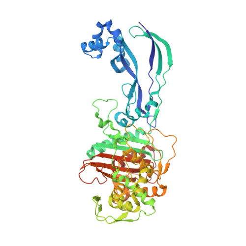
    Ceftobiprole is an advanced-generation broad-spectrum cephalosporin antibiotic with potent and rapid bactericidal activity against Gram-positive pathogens, including methicillin-resistant Staphylococcus aureus , as well as susceptible Gram-negative pathogens, including Pseudomonas sp. pathogens. In the case of Pseudomonas aeruginosa , ceftobiprole acts by inhibiting P. aeruginosa penicillin-binding protein 3 (PBP3). Structural studies were pursued to elucidate the molecular details of this PBP inhibition. The crystal structure of the His-tagged PBP3-ceftobiprole complex revealed a covalent bond between the ligand and the catalytic residue S294. Ceftobiprole binding leads to large active site changes near binding sites for the pyrrolidinone and pyrrolidine rings. The S528 to L536 region adopts a conformation previously not observed in PBP3, including partial unwinding of the α11 helix. These molecular insights can lead to a deeper understanding of β-lactam-PBP interactions that result in major changes in protein structure, as well as suggesting how to fine-tune current inhibitors and to develop novel inhibitors of this PBP.


  • Organizational Affiliation

    Department of Biochemistry, Case Western Reserve University, Cleveland, Ohio, USA.


Macromolecules
Find similar proteins by:  (by identity cutoff)  |  3D Structure
Entity ID: 1
MoleculeChains Sequence LengthOrganismDetailsImage
Peptidoglycan D,D-transpeptidase FtsI538Pseudomonas aeruginosaMutation(s): 0 
Gene Names: 
EC: 3.4.16.4
UniProt
Find proteins for G3XD46 (Pseudomonas aeruginosa (strain ATCC 15692 / DSM 22644 / CIP 104116 / JCM 14847 / LMG 12228 / 1C / PRS 101 / PAO1))
Explore G3XD46 
Go to UniProtKB:  G3XD46
Entity Groups  
Sequence Clusters30% Identity50% Identity70% Identity90% Identity95% Identity100% Identity
UniProt GroupG3XD46
Sequence Annotations
Expand
  • Reference Sequence
Small Molecules
Ligands 2 Unique
IDChains Name / Formula / InChI Key2D Diagram3D Interactions
RB6 (Subject of Investigation/LOI)
Query on RB6

Download Ideal Coordinates CCD File 
B [auth A](2R)-2-[(1R)-1-{[(2Z)-2-(5-amino-1,2,4-thiadiazol-3-yl)-2-(hydroxyimino)acetyl]amino}-2-oxoethyl]-5-({2-oxo-1-[(3R)-pyr rolidin-3-yl]-2,5-dihydro-1H-pyrrol-3-yl}methyl)-3,6-dihydro-2H-1,3-thiazine-4-carboxylic acid
C20 H24 N8 O6 S2
MYAXGJQBOYOEHQ-SWBIIUODSA-N
CL
Query on CL

Download Ideal Coordinates CCD File 
C [auth A],
D [auth A]
CHLORIDE ION
Cl
VEXZGXHMUGYJMC-UHFFFAOYSA-M
Experimental Data & Validation

Experimental Data

  • Method: X-RAY DIFFRACTION
  • Resolution: 1.76 Å
  • R-Value Free: 0.252 
  • R-Value Work: 0.205 
  • R-Value Observed: 0.207 
  • Space Group: P 21 21 21
Unit Cell:
Length ( Å )Angle ( ˚ )
a = 68.195α = 90
b = 83.739β = 90
c = 89.918γ = 90
Software Package:
Software NamePurpose
XDSdata reduction
REFMACrefinement
PDB_EXTRACTdata extraction
Aimlessdata scaling
PHASERphasing

Structure Validation

View Full Validation Report

Currently 6VJE does not have a validation slider image.


Entry History 

Deposition Data

Revision History  (Full details and data files)

  • Version 1.0: 2020-03-11
    Type: Initial release
  • Version 1.1: 2020-03-25
    Changes: Database references
  • Version 1.2: 2020-05-06
    Changes: Database references
  • Version 1.3: 2020-05-13
    Changes: Structure summary
  • Version 1.4: 2023-10-11
    Changes: Data collection, Database references, Derived calculations, Refinement description, Structure summary