6T6X

Structure of the Bottromycin epimerase BotH in complex with substrate


Experimental Data Snapshot

  • Method: X-RAY DIFFRACTION
  • Resolution: 1.25 Å
  • R-Value Free: 0.175 
  • R-Value Work: 0.166 
  • R-Value Observed: 0.167 

wwPDB Validation   3D Report Full Report


Ligand Structure Quality Assessment 


This is version 1.2 of the entry. See complete history


Literature

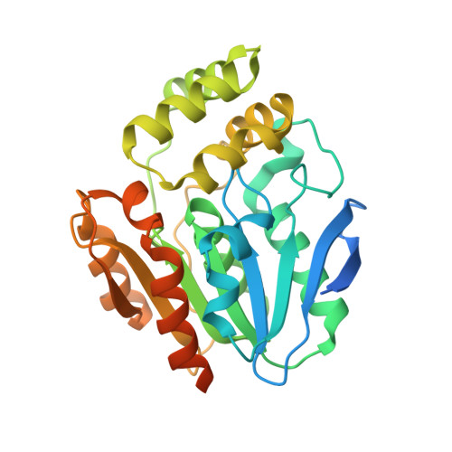
The bottromycin epimerase BotH defines a group of atypical alpha / beta-hydrolase-fold enzymes.

Sikandar, A.Franz, L.Adam, S.Santos-Aberturas, J.Horbal, L.Luzhetskyy, A.Truman, A.W.Kalinina, O.V.Koehnke, J.

(2020) Nat Chem Biol 16: 1013-1018

  • DOI: https://doi.org/10.1038/s41589-020-0569-y
  • Primary Citation of Related Structures:  
    6T6H, 6T6X, 6T6Y, 6T6Z, 6T70

  • PubMed Abstract: 

    D-amino acids endow peptides with diverse, desirable properties, but the post-translational and site-specific epimerization of L-amino acids into their D-counterparts is rare and chemically challenging. Bottromycins are ribosomally synthesized and post-translationally modified peptides that have overcome this challenge and feature a D-aspartate (D-Asp), which was proposed to arise spontaneously during biosynthesis. We have identified the highly unusual α/β-hydrolase (ABH) fold enzyme BotH as a peptide epimerase responsible for the post-translational epimerization of L-Asp to D-Asp during bottromycin biosynthesis. The biochemical characterization of BotH combined with the structures of BotH and the BotH-substrate complex allowed us to propose a mechanism for this reaction. Bioinformatic analyses of BotH homologs show that similar ABH enzymes are found in diverse biosynthetic gene clusters. This places BotH as the founding member of a group of atypical ABH enzymes that may be able to epimerize non-Asp stereocenters across different families of secondary metabolites.


  • Organizational Affiliation

    Workgroup Structural Biology of Biosynthetic Enzymes, Helmholtz Institute for Pharmaceutical Research Saarland (HIPS), Helmholtz Centre for Infection Research (HZI), Saarland University, Saarbrücken, Germany.


Macromolecules
Find similar proteins by:  (by identity cutoff)  |  3D Structure
Entity ID: 1
MoleculeChains Sequence LengthOrganismDetailsImage
BotH310Streptomyces sp. BC16019Mutation(s): 0 
Gene Names: botH
UniProt
Find proteins for K4MHV9 (Streptomyces sp. BC16019)
Explore K4MHV9 
Go to UniProtKB:  K4MHV9
Entity Groups  
Sequence Clusters30% Identity50% Identity70% Identity90% Identity95% Identity100% Identity
UniProt GroupK4MHV9
Sequence Annotations
Expand
  • Reference Sequence
Small Molecules
Ligands 1 Unique
IDChains Name / Formula / InChI Key2D Diagram3D Interactions
MQW (Subject of Investigation/LOI)
Query on MQW

Download Ideal Coordinates CCD File 
B [auth A](4~{R})-2-[(1~{R})-1-[[(2~{S})-2-[[(2~{S})-3-methyl-2-[[(4~{Z},6~{S},9~{S},12~{S})-2,8,11-tris(oxidanylidene)-6,9-di(propan-2-yl)-1,4,7,10-tetrazabicyclo[10.3.0]pentadec-4-en-5-yl]amino]butanoyl]amino]-3-phenyl-propanoyl]amino]-3-oxidanyl-3-oxidanylidene-propyl]-4,5-dihydro-1,3-thiazole-4-carboxylic acid
C38 H54 N8 O9 S
PXXAKUWJCGKVNK-JQXGNVTOSA-N
Experimental Data & Validation

Experimental Data

  • Method: X-RAY DIFFRACTION
  • Resolution: 1.25 Å
  • R-Value Free: 0.175 
  • R-Value Work: 0.166 
  • R-Value Observed: 0.167 
  • Space Group: I 2 2 2
Unit Cell:
Length ( Å )Angle ( ˚ )
a = 66.655α = 90
b = 80.285β = 90
c = 89.337γ = 90
Software Package:
Software NamePurpose
PHENIXrefinement
XDSdata reduction
SCALAdata scaling
PHASERphasing

Structure Validation

View Full Validation Report


Ligand Structure Quality Assessment 


Entry History & Funding Information

Deposition Data


Funding OrganizationLocationGrant Number
German Research FoundationGermanyKO 4116/3-1

Revision History  (Full details and data files)

  • Version 1.0: 2020-07-15
    Type: Initial release
  • Version 1.1: 2020-08-26
    Changes: Database references
  • Version 1.2: 2024-01-24
    Changes: Data collection, Database references, Refinement description