6RT8

Structure of catharanthine synthase - an alpha-beta hydrolase from Catharanthus roseus with a cleaviminium intermediate bound


Experimental Data Snapshot

  • Method: X-RAY DIFFRACTION
  • Resolution: 2.19 Å
  • R-Value Free: 0.234 
  • R-Value Work: 0.194 
  • R-Value Observed: 0.196 

wwPDB Validation   3D Report Full Report


Ligand Structure Quality Assessment 


This is version 1.2 of the entry. See complete history


Literature

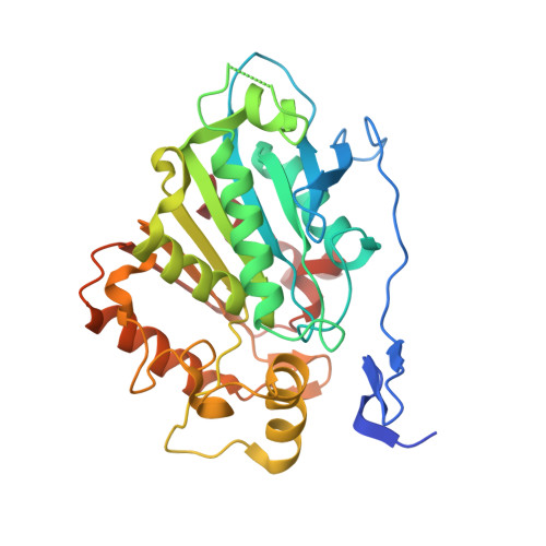
Structural basis of cycloaddition in biosynthesis of iboga and aspidosperma alkaloids.

Caputi, L.Franke, J.Bussey, K.Farrow, S.C.Vieira, I.J.C.Stevenson, C.E.M.Lawson, D.M.O'Connor, S.E.

(2020) Nat Chem Biol 16: 383-386

  • DOI: https://doi.org/10.1038/s41589-019-0460-x
  • Primary Citation of Related Structures:  
    6RJ8, 6RS4, 6RT8

  • PubMed Abstract: 

    Cycloaddition reactions generate chemical complexity in a single step. Here we report the crystal structures of three homologous plant-derived cyclases involved in the biosynthesis of iboga and aspidosperma alkaloids. These enzymes act on the same substrate, named angryline, to generate three distinct scaffolds. Mutational analysis reveals how these highly similar enzymes control regio- and stereo-selectivity.


  • Organizational Affiliation

    Max Planck Institute of Chemical Ecology, Department of Natural Product Biosynthesis, Jena, Germany.


Macromolecules
Find similar proteins by:  (by identity cutoff)  |  3D Structure
Entity ID: 1
MoleculeChains Sequence LengthOrganismDetailsImage
Catharanthine synthase
A, B, C, D, E
A, B, C, D, E, F, G, H
330Catharanthus roseusMutation(s): 0 
Gene Names: CSHL1Caros025416
EC: 4
UniProt
Find proteins for A0A2P1GIW2 (Catharanthus roseus)
Explore A0A2P1GIW2 
Go to UniProtKB:  A0A2P1GIW2
Entity Groups  
Sequence Clusters30% Identity50% Identity70% Identity90% Identity95% Identity100% Identity
UniProt GroupA0A2P1GIW2
Sequence Annotations
Expand
  • Reference Sequence
Small Molecules
Ligands 2 Unique
IDChains Name / Formula / InChI Key2D Diagram3D Interactions
KJE (Subject of Investigation/LOI)
Query on KJE

Download Ideal Coordinates CCD File 
I [auth A]
K [auth B]
L [auth C]
N [auth D]
O [auth E]
I [auth A],
K [auth B],
L [auth C],
N [auth D],
O [auth E],
Q [auth F],
R [auth G],
S [auth H]
18-carboxymethoxy-cleaviminium
C21 H25 N2 O2
ULODILYUCKTWMV-CGTJXYLNSA-N
P6G
Query on P6G

Download Ideal Coordinates CCD File 
J [auth A],
M [auth C],
P [auth E],
T [auth H]
HEXAETHYLENE GLYCOL
C12 H26 O7
IIRDTKBZINWQAW-UHFFFAOYSA-N
Experimental Data & Validation

Experimental Data

  • Method: X-RAY DIFFRACTION
  • Resolution: 2.19 Å
  • R-Value Free: 0.234 
  • R-Value Work: 0.194 
  • R-Value Observed: 0.196 
  • Space Group: P 1 21 1
Unit Cell:
Length ( Å )Angle ( ˚ )
a = 81.54α = 90
b = 121.03β = 99.02
c = 157.98γ = 90
Software Package:
Software NamePurpose
XDSdata reduction
Aimlessdata scaling
REFMACrefinement
PDB_EXTRACTdata extraction
PHASERphasing

Structure Validation

View Full Validation Report


Ligand Structure Quality Assessment 


Entry History & Funding Information

Deposition Data


Funding OrganizationLocationGrant Number
European Research CouncilBelgium788301
Biotechnology and Biological Sciences Research CouncilUnited KingdomBB/P012523/1

Revision History  (Full details and data files)

  • Version 1.0: 2020-03-04
    Type: Initial release
  • Version 1.1: 2020-04-01
    Changes: Database references
  • Version 1.2: 2024-01-24
    Changes: Data collection, Database references, Refinement description