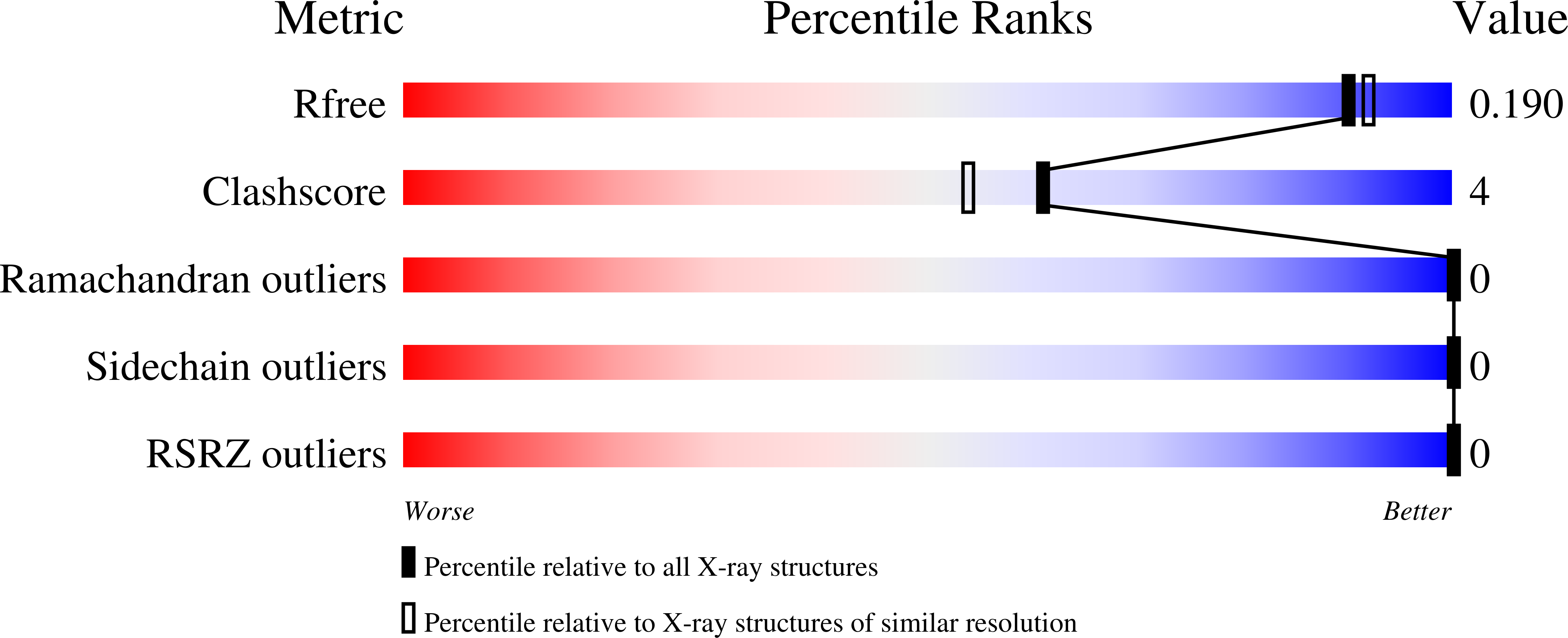
6R35

Structure of the LecB lectin from Pseudomonas aeruginosa strain PAO1 in complex with lewis x tetrasaccharide


Experimental Data Snapshot

  • Method: X-RAY DIFFRACTION
  • Resolution: 1.80 Å
  • R-Value Free: 0.176 
  • R-Value Work: 0.132 
  • R-Value Observed: 0.134 

wwPDB Validation   3D Report Full Report


This is version 2.1 of the entry. See complete history


Literature

Induction of rare conformation of oligosaccharide by binding to calcium-dependent bacterial lectin: X-ray crystallography and modelling study.

Lepsik, M.Sommer, R.Kuhaudomlarp, S.Lelimousin, M.Paci, E.Varrot, A.Titz, A.Imberty, A.

(2019) Eur J Med Chem 177: 212-220

  • DOI: https://doi.org/10.1016/j.ejmech.2019.05.049
  • Primary Citation of Related Structures:  
    5A70, 6R35

  • PubMed Abstract: 

    Pathogenic micro-organisms utilize protein receptors (lectins) in adhesion to host tissues, a process that in some cases relies on the interaction between lectins and human glycoconjugates. Oligosaccharide epitopes are recognized through their three-dimensional structure and their flexibility is a key issue in specificity. In this paper, we analysed by X-ray crystallography the structures of the LecB lectin from two strains of Pseudomonas aeruginosa in complex with Lewis x oligosaccharide present on cell surfaces of human tissues. An unusual conformation of the glycan was observed in all binding sites with a non-canonical syn orientation of the N-acetyl group of N-acetyl-glucosamine. A PDB-wide search revealed that such an orientation occurs only in 4% of protein/carbohydrate complexes. Theoretical chemistry calculations showed that the observed conformation is unstable in solution but stabilised by the lectin. A reliable description of LecB/Lewis x complex by force field-based methods had proven especially challenging due to the special feature of the binding site, two closely apposed Ca 2+ ions which induce strong charge delocalisation. By comparing various force-field parametrisations, we propose a general strategy which will be useful in near future for designing carbohydrate-based ligands (glycodrugs) against other calcium-dependent protein receptors.


  • Organizational Affiliation

    Université Grenoble Alpes, CNRS, CERMAV, 38000, Grenoble, France. Electronic address: martin.lepsik@cermav.cnrs.fr.


Macromolecules
Find similar proteins by:  (by identity cutoff)  |  3D Structure
Entity ID: 1
MoleculeChains Sequence LengthOrganismDetailsImage
Fucose-binding lectin PA-IILA [auth D],
B [auth A],
C [auth B],
D [auth C]
114Pseudomonas aeruginosa PAO1Mutation(s): 0 
Gene Names: lecBPA3361
UniProt
Find proteins for Q9HYN5 (Pseudomonas aeruginosa (strain ATCC 15692 / DSM 22644 / CIP 104116 / JCM 14847 / LMG 12228 / 1C / PRS 101 / PAO1))
Explore Q9HYN5 
Go to UniProtKB:  Q9HYN5
Entity Groups  
Sequence Clusters30% Identity50% Identity70% Identity90% Identity95% Identity100% Identity
UniProt GroupQ9HYN5
Sequence Annotations
Expand
  • Reference Sequence
Oligosaccharides

Help

Entity ID: 2
MoleculeChains Length2D Diagram Glycosylation3D Interactions
alpha-L-fucopyranose-(1-3)-[beta-D-galactopyranose-(1-4)]2-acetamido-2-deoxy-beta-D-glucopyranose
E, F, G, H
3N/A
Glycosylation Resources
GlyTouCan:  G00051MO
GlyCosmos:  G00051MO
GlyGen:  G00051MO
Small Molecules
Ligands 3 Unique
IDChains Name / Formula / InChI Key2D Diagram3D Interactions
SO4
Query on SO4

Download Ideal Coordinates CCD File 
K [auth D],
W [auth C]
SULFATE ION
O4 S
QAOWNCQODCNURD-UHFFFAOYSA-L
EDO
Query on EDO

Download Ideal Coordinates CCD File 
L [auth D]
P [auth A]
Q [auth A]
T [auth B]
U [auth B]
L [auth D],
P [auth A],
Q [auth A],
T [auth B],
U [auth B],
X [auth C],
Y [auth C],
Z [auth C]
1,2-ETHANEDIOL
C2 H6 O2
LYCAIKOWRPUZTN-UHFFFAOYSA-N
CA (Subject of Investigation/LOI)
Query on CA

Download Ideal Coordinates CCD File 
I [auth D]
J [auth D]
M [auth D]
N [auth A]
O [auth A]
I [auth D],
J [auth D],
M [auth D],
N [auth A],
O [auth A],
R [auth A],
S [auth B],
V [auth C]
CALCIUM ION
Ca
BHPQYMZQTOCNFJ-UHFFFAOYSA-N
Experimental Data & Validation

Experimental Data

  • Method: X-RAY DIFFRACTION
  • Resolution: 1.80 Å
  • R-Value Free: 0.176 
  • R-Value Work: 0.132 
  • R-Value Observed: 0.134 
  • Space Group: P 1 21 1
Unit Cell:
Length ( Å )Angle ( ˚ )
a = 52.603α = 90
b = 72.481β = 114.62
c = 62.061γ = 90
Software Package:
Software NamePurpose
XDSdata reduction
Aimlessdata scaling
REFMACrefinement
PDB_EXTRACTdata extraction
PHASERphasing

Structure Validation

View Full Validation Report


Entry History & Funding Information

Deposition Data


Funding OrganizationLocationGrant Number
European UnionFrance795605

Revision History  (Full details and data files)

  • Version 1.0: 2019-06-12
    Type: Initial release
  • Version 1.1: 2019-09-18
    Changes: Data collection, Refinement description
  • Version 2.0: 2020-07-29
    Type: Remediation
    Reason: Carbohydrate remediation
    Changes: Atomic model, Data collection, Derived calculations, Structure summary
  • Version 2.1: 2024-01-24
    Changes: Data collection, Database references, Refinement description, Structure summary