Structural insights into dehydratase substrate selection for the borrelidin and fluvirucin polyketide synthases.
Barajas, J.F., McAndrew, R.P., Thompson, M.G., Backman, T.W.H., Pang, B., de Rond, T., Pereira, J.H., Benites, V.T., Martin, H.G., Baidoo, E.E.K., Hillson, N.J., Adams, P.D., Keasling, J.D.(2019) J Ind Microbiol Biotechnol 46: 1225-1235
- PubMed: 31115703
- DOI: https://doi.org/10.1007/s10295-019-02189-z
- Primary Citation of Related Structures:
6OBT, 6OBV - PubMed Abstract:
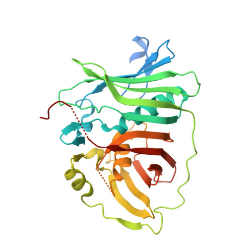
Engineered polyketide synthases (PKSs) are promising synthetic biology platforms for the production of chemicals with diverse applications. The dehydratase (DH) domain within modular type I PKSs generates an α,β-unsaturated bond in nascent polyketide intermediates through a dehydration reaction. Several crystal structures of DH domains have been solved, providing important structural insights into substrate selection and dehydration. Here, we present two DH domain structures from two chemically diverse PKSs. The first DH domain, isolated from the third module in the borrelidin PKS, is specific towards a trans-cyclopentane-carboxylate-containing polyketide substrate. The second DH domain, isolated from the first module in the fluvirucin B 1 PKS, accepts an amide-containing polyketide intermediate. Sequence-structure analysis of these domains, in addition to previously published DH structures, display many significant similarities and key differences pertaining to substrate selection. The two major differences between BorA DH M3, FluA DH M1 and other DH domains are found in regions of unmodeled residues or residues containing high B-factors. These two regions are located between α3-β11 and β7-α2. From the catalytic Asp located in α3 to a conserved Pro in β11, the residues between them form part of the bottom of the substrate-binding cavity responsible for binding to acyl-ACP intermediates.
Organizational Affiliation:
Department of Energy Agile BioFoundry, Emeryville, CA, 94608, USA.