6NIZ

Atomic structure of a fluorescent Ag8 cluster templated by a multistranded DNA scaffold


Experimental Data Snapshot

  • Method: X-RAY DIFFRACTION
  • Resolution: 0.93 Å
  • R-Value Free: 0.091 
  • R-Value Work: 0.082 
  • R-Value Observed: 0.083 

wwPDB Validation   3D Report Full Report


This is version 1.4 of the entry. See complete history


Literature

Atomic Structure of a Fluorescent Ag8Cluster Templated by a Multistranded DNA Scaffold.

Huard, D.J.E.Demissie, A.Kim, D.Lewis, D.Dickson, R.M.Petty, J.T.Lieberman, R.L.

(2019) J Am Chem Soc 141: 11465-11470

  • DOI: https://doi.org/10.1021/jacs.8b12203
  • Primary Citation of Related Structures:  
    6NIZ

  • PubMed Abstract: 

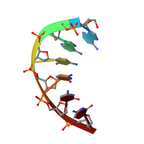
    Multinuclear silver clusters encapsulated by DNA exhibit size-tunable emission spectra and rich photophysics, but their atomic organization is poorly understood. Herein, we describe the structure of one such hybrid chromophore, a green-emitting Ag 8 cluster arranged in a Big Dipper-shape bound to the oligonucleotide A 2 C 4 . Three 3' cytosine metallo-base pairs stabilize a parallel A-form-like duplex with a 5' adenine-rich pocket, which binds a metallic, trapezoidal-shaped Ag 5 moiety via Ag-N bonds to endo- and exocyclic nitrogens of cytosine and adenine. The unique DNA configuration, constrained coordination environment, and templated Ag 8 cluster arrangement highlight the reciprocity between the silvers and DNA in adopting this structure. These first atomic details of a DNA-encapsulated Ag cluster fluorophore illuminate many aspects of biological assembly, nanoscience, and metal cluster photophysics.


  • Organizational Affiliation

    School of Chemistry & Biochemistry and Parker H. Petit Institute for Bioengineering and Bioscience , Georgia Institute of Technology , Atlanta , Georgia 30332 , United States.


Macromolecules

Find similar nucleic acids by:  Sequence   |   3D Structure  

Entity ID: 1
MoleculeChains LengthOrganismImage
DNA (5'-D(*AP*AP*CP*CP*CP*CP)-3')
A, B
6synthetic construct
Sequence Annotations
Expand
  • Reference Sequence
Small Molecules
Ligands 1 Unique
IDChains Name / Formula / InChI Key2D Diagram3D Interactions
AG
Query on AG

Download Ideal Coordinates CCD File 
C [auth A]
D [auth A]
E [auth A]
F [auth A]
G [auth A]
C [auth A],
D [auth A],
E [auth A],
F [auth A],
G [auth A],
H [auth A],
I [auth A],
J [auth B],
K [auth B],
L [auth B],
M [auth B],
N [auth B]
SILVER ION
Ag
FOIXSVOLVBLSDH-UHFFFAOYSA-N
Experimental Data & Validation

Experimental Data

  • Method: X-RAY DIFFRACTION
  • Resolution: 0.93 Å
  • R-Value Free: 0.091 
  • R-Value Work: 0.082 
  • R-Value Observed: 0.083 
  • Space Group: P 41 21 2
Unit Cell:
Length ( Å )Angle ( ˚ )
a = 33.29α = 90
b = 33.29β = 90
c = 62.01γ = 90
Software Package:
Software NamePurpose
PHENIXrefinement
XDSdata reduction
XSCALEdata scaling
PHASERphasing

Structure Validation

View Full Validation Report


Entry History & Funding Information

Deposition Data


Funding OrganizationLocationGrant Number
National Science Foundation (NSF, United States)United States1817796

Revision History  (Full details and data files)

  • Version 1.0: 2019-01-16
    Type: Initial release
  • Version 1.1: 2019-06-26
    Changes: Data collection, Database references
  • Version 1.2: 2019-07-31
    Changes: Data collection, Database references
  • Version 1.3: 2019-11-27
    Changes: Author supporting evidence
  • Version 1.4: 2024-03-13
    Changes: Data collection, Database references, Derived calculations