6K3C

Crystal structure of class I PHA synthase (PhaC) mutant from Chromobacterium sp. USM2 bound to Coenzyme A.


Experimental Data Snapshot

  • Method: X-RAY DIFFRACTION
  • Resolution: 3.07 Å
  • R-Value Free: 0.263 
  • R-Value Work: 0.224 
  • R-Value Observed: 0.226 

wwPDB Validation   3D Report Full Report


Ligand Structure Quality Assessment 


This is version 1.2 of the entry. See complete history


Literature

Asymmetric Open-Closed Dimer Mechanism of Polyhydroxyalkanoate Synthase PhaC.

Chek, M.F.Kim, S.Y.Mori, T.Tan, H.T.Sudesh, K.Hakoshima, T.

(2020) iScience 23: 101084-101084

  • DOI: https://doi.org/10.1016/j.isci.2020.101084
  • Primary Citation of Related Structures:  
    6K3C

  • PubMed Abstract: 

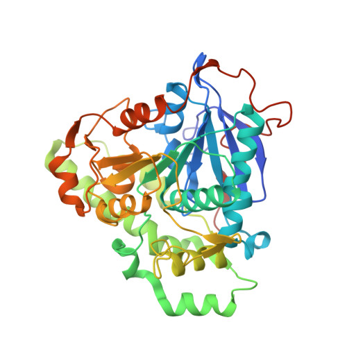
    Biodegradable polyester polyhydroxyalkanoate (PHA) is a promising bioplastic material for industrial use as a replacement for petroleum-based plastics. PHA synthase PhaC forms an active dimer to polymerize acyl moieties from the substrate acyl-coenzyme A (CoA) into PHA polymers. Here we present the crystal structure of the catalytic domain of PhaC from Chromobacterium sp. USM2, bound to CoA. The structure reveals an asymmetric dimer, in which one protomer adopts an open conformation bound to CoA, whereas the other adopts a closed conformation in a CoA-free form. The open conformation is stabilized by the asymmetric dimerization and enables PhaC to accommodate CoA and also to create the product egress path. The bound CoA molecule has its β-mercaptoethanolamine moiety extended into the active site with the terminal SH group close to active center Cys291, enabling formation of the reaction intermediate by acylation of Cys291.


  • Organizational Affiliation

    Structural Biology Laboratory, Nara Institute of Science and Technology, 8916-5 Takayama, Ikoma, Nara 630-0192, Japan.


Macromolecules
Find similar proteins by:  (by identity cutoff)  |  3D Structure
Entity ID: 1
MoleculeChains Sequence LengthOrganismDetailsImage
Intracellular polyhydroxyalkanoate synthase
A, B
395Chromobacterium sp. USM2Mutation(s): 1 
Gene Names: phaC
UniProt
Find proteins for E1APK1 (Chromobacterium sp. USM2)
Explore E1APK1 
Go to UniProtKB:  E1APK1
Entity Groups  
Sequence Clusters30% Identity50% Identity70% Identity90% Identity95% Identity100% Identity
UniProt GroupE1APK1
Sequence Annotations
Expand
  • Reference Sequence
Small Molecules
Ligands 1 Unique
IDChains Name / Formula / InChI Key2D Diagram3D Interactions
COA (Subject of Investigation/LOI)
Query on COA

Download Ideal Coordinates CCD File 
C [auth B]COENZYME A
C21 H36 N7 O16 P3 S
RGJOEKWQDUBAIZ-IBOSZNHHSA-N
Experimental Data & Validation

Experimental Data

  • Method: X-RAY DIFFRACTION
  • Resolution: 3.07 Å
  • R-Value Free: 0.263 
  • R-Value Work: 0.224 
  • R-Value Observed: 0.226 
  • Space Group: C 1 2 1
Unit Cell:
Length ( Å )Angle ( ˚ )
a = 169.802α = 90
b = 61.852β = 106.73
c = 82.566γ = 90
Software Package:
Software NamePurpose
PHENIXrefinement
HKL-2000data reduction
HKL-2000data scaling
PHENIXphasing

Structure Validation

View Full Validation Report


Ligand Structure Quality Assessment 


Entry History 

Deposition Data

Revision History  (Full details and data files)

  • Version 1.0: 2020-04-29
    Type: Initial release
  • Version 1.1: 2020-05-27
    Changes: Database references
  • Version 1.2: 2023-11-22
    Changes: Data collection, Database references, Refinement description