6JIB

Human MTHFD2 in complex with DS44960156


Experimental Data Snapshot

  • Method: X-RAY DIFFRACTION
  • Resolution: 2.25 Å
  • R-Value Free: 0.243 
  • R-Value Work: 0.201 
  • R-Value Observed: 0.203 

wwPDB Validation   3D Report Full Report


Ligand Structure Quality Assessment 


This is version 1.2 of the entry. See complete history


Literature

Structure-Based Design and Synthesis of an Isozyme-Selective MTHFD2 Inhibitor with a Tricyclic Coumarin Scaffold.

Kawai, J.Ota, M.Ohki, H.Toki, T.Suzuki, M.Shimada, T.Matsui, S.Inoue, H.Sugihara, C.Matsuhashi, N.Matsui, Y.Takaishi, S.Nakayama, K.

(2019) ACS Med Chem Lett 10: 893-898

  • DOI: https://doi.org/10.1021/acsmedchemlett.9b00069
  • Primary Citation of Related Structures:  
    6JIB, 6JID

  • PubMed Abstract: 

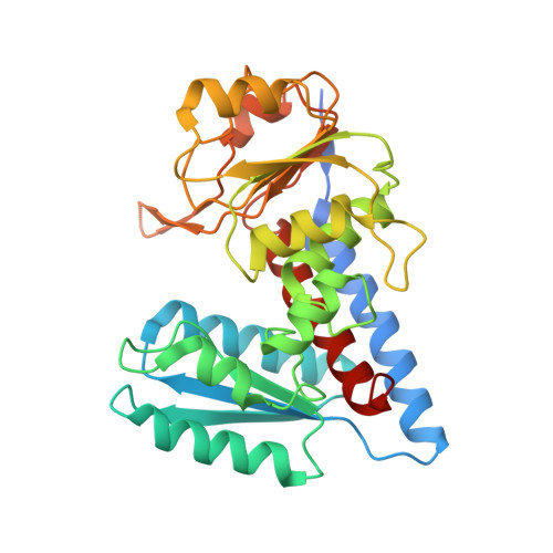
    Methylenetetrahydrofolate dehydrogenase 2 (MTHFD2) plays a key role in one-carbon (1C) metabolism in human mitochondria, and its high expression correlates with poor survival of patients with various types of cancer. An isozyme-selective MTHFD2 inhibitor is highly attractive for potential use in cancer treatment. Herein, we disclose a novel isozyme-selective MTHFD2 inhibitor DS44960156, with a tricyclic coumarin scaffold, which was initially discovered via high-throughput screening (HTS) and improved using structure-based drug design (SBDD). DS44960156 would offer a good starting point for further optimization based on the following features: (1) unprecedented selectivity (>18-fold) for MTHFD2 over MTHFD1, (2) a molecular weight of less than 400, and (3) good ligand efficiency (LE).


  • Organizational Affiliation

    R&D Division, Daiichi Sankyo Co., Ltd., 1-2-58 Hiromachi, Shinagawa-ku, Tokyo 140-8710, Japan.


Macromolecules
Find similar proteins by:  (by identity cutoff)  |  3D Structure
Entity ID: 1
MoleculeChains Sequence LengthOrganismDetailsImage
Bifunctional methylenetetrahydrofolate dehydrogenase/cyclohydrolase, mitochondrial
A, B
323Homo sapiensMutation(s): 0 
Gene Names: MTHFD2NMDMC
EC: 1.5.1.15 (PDB Primary Data), 3.5.4.9 (PDB Primary Data)
UniProt & NIH Common Fund Data Resources
Find proteins for P13995 (Homo sapiens)
Explore P13995 
Go to UniProtKB:  P13995
PHAROS:  P13995
GTEx:  ENSG00000065911 
Entity Groups  
Sequence Clusters30% Identity50% Identity70% Identity90% Identity95% Identity100% Identity
UniProt GroupP13995
Sequence Annotations
Expand
  • Reference Sequence
Small Molecules
Ligands 3 Unique
IDChains Name / Formula / InChI Key2D Diagram3D Interactions
BQF (Subject of Investigation/LOI)
Query on BQF

Download Ideal Coordinates CCD File 
C [auth A],
E [auth A],
I [auth B],
J [auth B]
4-(5-oxo-1,5-dihydro-2H-[1]benzopyrano[3,4-c]pyridine-3(4H)-carbonyl)benzoic acid
C20 H15 N O5
OXUKTNZACAKVRG-UHFFFAOYSA-N
PO4
Query on PO4

Download Ideal Coordinates CCD File 
D [auth A],
H [auth A]
PHOSPHATE ION
O4 P
NBIIXXVUZAFLBC-UHFFFAOYSA-K
GOL
Query on GOL

Download Ideal Coordinates CCD File 
F [auth A],
G [auth A],
K [auth B],
L [auth B],
M [auth B]
GLYCEROL
C3 H8 O3
PEDCQBHIVMGVHV-UHFFFAOYSA-N
Binding Affinity Annotations 
IDSourceBinding Affinity
BQF Binding MOAD:  6JIB IC50: 1600 (nM) from 1 assay(s)
Experimental Data & Validation

Experimental Data

  • Method: X-RAY DIFFRACTION
  • Resolution: 2.25 Å
  • R-Value Free: 0.243 
  • R-Value Work: 0.201 
  • R-Value Observed: 0.203 
  • Space Group: P 65
Unit Cell:
Length ( Å )Angle ( ˚ )
a = 116.424α = 90
b = 116.424β = 90
c = 113.178γ = 120
Software Package:
Software NamePurpose
XDSdata reduction
Aimlessdata scaling
REFMACrefinement
PDB_EXTRACTdata extraction
PHASERphasing

Structure Validation

View Full Validation Report


Ligand Structure Quality Assessment 


Entry History 

Deposition Data

Revision History  (Full details and data files)

  • Version 1.0: 2019-06-05
    Type: Initial release
  • Version 1.1: 2019-07-10
    Changes: Data collection, Database references
  • Version 1.2: 2023-11-22
    Changes: Data collection, Database references, Refinement description