6I7G

Crystal structure of dimeric wild type FICD complexed with ATP


Experimental Data Snapshot

  • Method: X-RAY DIFFRACTION
  • Resolution: 2.70 Å
  • R-Value Free: 0.319 
  • R-Value Work: 0.283 
  • R-Value Observed: 0.285 

wwPDB Validation   3D Report Full Report


Ligand Structure Quality Assessment 


This is version 1.3 of the entry. See complete history


Literature

An oligomeric state-dependent switch in the ER enzyme FICD regulates AMPylation and deAMPylation of BiP.

Perera, L.A.Rato, C.Yan, Y.Neidhardt, L.McLaughlin, S.H.Read, R.J.Preissler, S.Ron, D.

(2019) EMBO J 38: e102177-e102177

  • DOI: https://doi.org/10.15252/embj.2019102177
  • Primary Citation of Related Structures:  
    6I7G, 6I7H, 6I7I, 6I7J, 6I7K, 6I7L

  • PubMed Abstract: 

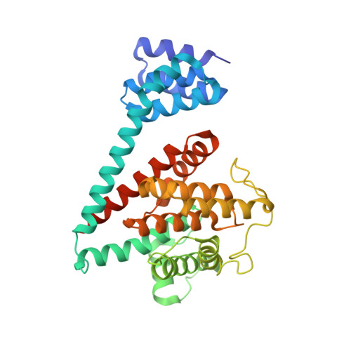
    AMPylation is an inactivating modification that alters the activity of the major endoplasmic reticulum (ER) chaperone BiP to match the burden of unfolded proteins. A single ER-localised Fic protein, FICD (HYPE), catalyses both AMPylation and deAMPylation of BiP. However, the basis for the switch in FICD's activity is unknown. We report on the transition of FICD from a dimeric enzyme, that deAMPylates BiP, to a monomer with potent AMPylation activity. Mutations in the dimer interface, or of residues along an inhibitory pathway linking the dimer interface to the enzyme's active site, favour BiP AMPylation in vitro and in cells. Mechanistically, monomerisation relieves a repressive effect allosterically propagated from the dimer interface to the inhibitory Glu234, thereby permitting AMPylation-competent binding of MgATP. Moreover, a reciprocal signal, propagated from the nucleotide-binding site, provides a mechanism for coupling the oligomeric state and enzymatic activity of FICD to the energy status of the ER.


  • Organizational Affiliation

    Cambridge Institute for Medical Research, University of Cambridge, Cambridge, UK.


Macromolecules
Find similar proteins by:  (by identity cutoff)  |  3D Structure
Entity ID: 1
MoleculeChains Sequence LengthOrganismDetailsImage
Adenosine monophosphate-protein transferase FICD
A, B
343Homo sapiensMutation(s): 0 
Gene Names: FICDHIP13HYPEUNQ3041/PRO9857
EC: 2.7.7 (PDB Primary Data), 3.1.4 (PDB Primary Data)
UniProt & NIH Common Fund Data Resources
Find proteins for Q9BVA6 (Homo sapiens)
Explore Q9BVA6 
Go to UniProtKB:  Q9BVA6
PHAROS:  Q9BVA6
GTEx:  ENSG00000198855 
Entity Groups  
Sequence Clusters30% Identity50% Identity70% Identity90% Identity95% Identity100% Identity
UniProt GroupQ9BVA6
Sequence Annotations
Expand
  • Reference Sequence
Small Molecules
Experimental Data & Validation

Experimental Data

  • Method: X-RAY DIFFRACTION
  • Resolution: 2.70 Å
  • R-Value Free: 0.319 
  • R-Value Work: 0.283 
  • R-Value Observed: 0.285 
  • Space Group: P 21 21 2
Unit Cell:
Length ( Å )Angle ( ˚ )
a = 77.671α = 90
b = 107.652β = 90
c = 132.597γ = 90
Software Package:
Software NamePurpose
REFMACrefinement
xia2data reduction
Aimlessdata scaling
PHASERphasing

Structure Validation

View Full Validation Report


Ligand Structure Quality Assessment 


Entry History & Funding Information

Deposition Data


Funding OrganizationLocationGrant Number
Medical Research Council (United Kingdom)United KingdomMR/K50127X/1
Wellcome TrustUnited Kingdom200848/Z/16/Z
Wellcome TrustUnited Kingdom082961/Z/07/Z
Wellcome TrustUnited Kingdom100140

Revision History  (Full details and data files)

  • Version 1.0: 2019-09-25
    Type: Initial release
  • Version 1.1: 2019-10-02
    Changes: Data collection, Database references
  • Version 1.2: 2019-11-13
    Changes: Database references
  • Version 1.3: 2024-01-24
    Changes: Data collection, Database references, Refinement description