6I53

Cryo-EM structure of the human synaptic alpha1-beta3-gamma2 GABAA receptor in complex with Megabody38 in a lipid nanodisc


Experimental Data Snapshot

  • Method: ELECTRON MICROSCOPY
  • Resolution: 3.20 Å
  • Aggregation State: PARTICLE 
  • Reconstruction Method: SINGLE PARTICLE 

wwPDB Validation   3D Report Full Report


This is version 2.1 of the entry. See complete history


Literature

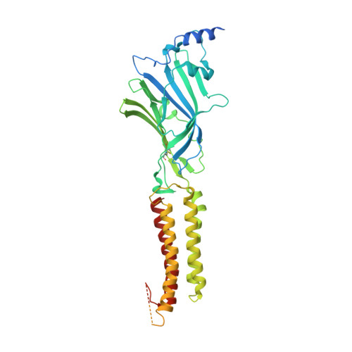
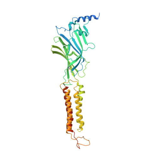
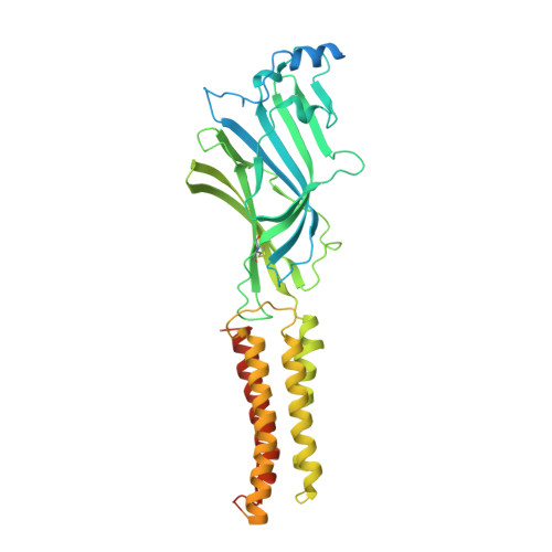
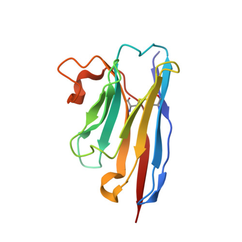
Cryo-EM structure of the human alpha 1 beta 3 gamma 2 GABAAreceptor in a lipid bilayer.

Laverty, D.Desai, R.Uchanski, T.Masiulis, S.Stec, W.J.Malinauskas, T.Zivanov, J.Pardon, E.Steyaert, J.Miller, K.W.Aricescu, A.R.

(2019) Nature 565: 516-520

  • DOI: https://doi.org/10.1038/s41586-018-0833-4
  • Primary Citation of Related Structures:  
    6I53

  • PubMed Abstract: 

    Type A γ-aminobutyric acid (GABA A ) receptors are pentameric ligand-gated ion channels and the main drivers of fast inhibitory neurotransmission in the vertebrate nervous system 1,2 . Their dysfunction is implicated in a range of neurological disorders, including depression, epilepsy and schizophrenia 3,4 . Among the numerous assemblies that are theoretically possible, the most prevalent in the brain are the α1β2/3γ2 GABA A receptors 5 . The β3 subunit has an important role in maintaining inhibitory tone, and the expression of this subunit alone is sufficient to rescue inhibitory synaptic transmission in β1-β3 triple knockout neurons 6 . So far, efforts to generate accurate structural models for heteromeric GABA A receptors have been hampered by the use of engineered receptors and the presence of detergents 7-9 . Notably, some recent cryo-electron microscopy reconstructions have reported 'collapsed' conformations 8,9 ; however, these disagree with the structure of the prototypical pentameric ligand-gated ion channel the Torpedo nicotinic acetylcholine receptor 10,11 , the large body of structural work on homologous homopentameric receptor variants 12 and the logic of an ion-channel architecture. Here we present a high-resolution cryo-electron microscopy structure of the full-length human α1β3γ2L-a major synaptic GABA A receptor isoform-that is functionally reconstituted in lipid nanodiscs. The receptor is bound to a positive allosteric modulator 'megabody' and is in a desensitized conformation. Each GABA A receptor pentamer contains two phosphatidylinositol-4,5-bisphosphate molecules, the head groups of which occupy positively charged pockets in the intracellular juxtamembrane regions of α1 subunits. Beyond this level, the intracellular M3-M4 loops are largely disordered, possibly because interacting post-synaptic proteins are not present. This structure illustrates the molecular principles of heteromeric GABA A receptor organization and provides a reference framework for future mechanistic investigations of GABAergic signalling and pharmacology.


  • Organizational Affiliation

    MRC Laboratory of Molecular Biology, Cambridge Biomedical Campus, Cambridge, UK. dlaverty@mrc-lmb.cam.ac.uk.


Macromolecules
Find similar proteins by:  (by identity cutoff)  |  3D Structure
Entity ID: 1
MoleculeChains Sequence LengthOrganismDetailsImage
Gamma-aminobutyric acid receptor subunit beta-3A [auth E],
C [auth B]
473Homo sapiensMutation(s): 0 
Gene Names: GABRB3
Membrane Entity: Yes 
UniProt & NIH Common Fund Data Resources
Find proteins for P28472 (Homo sapiens)
Explore P28472 
Go to UniProtKB:  P28472
PHAROS:  P28472
GTEx:  ENSG00000166206 
Entity Groups  
Sequence Clusters30% Identity50% Identity70% Identity90% Identity95% Identity100% Identity
UniProt GroupP28472
Sequence Annotations
Expand
  • Reference Sequence
Find similar proteins by:  (by identity cutoff)  |  3D Structure
Entity ID: 2
MoleculeChains Sequence LengthOrganismDetailsImage
Gamma-aminobutyric acid receptor subunit alpha-1B [auth A],
E [auth D]
464Homo sapiensMutation(s): 0 
Gene Names: GABRA1
Membrane Entity: Yes 
UniProt & NIH Common Fund Data Resources
Find proteins for P14867 (Homo sapiens)
Explore P14867 
Go to UniProtKB:  P14867
PHAROS:  P14867
GTEx:  ENSG00000022355 
Entity Groups  
Sequence Clusters30% Identity50% Identity70% Identity90% Identity95% Identity100% Identity
UniProt GroupP14867
Sequence Annotations
Expand
  • Reference Sequence
Find similar proteins by:  (by identity cutoff)  |  3D Structure
Entity ID: 3
MoleculeChains Sequence LengthOrganismDetailsImage
Gamma-aminobutyric acid receptor subunit gamma-2D [auth C]495Homo sapiensMutation(s): 0 
Gene Names: GABRG2
Membrane Entity: Yes 
UniProt & NIH Common Fund Data Resources
Find proteins for P18507 (Homo sapiens)
Explore P18507 
Go to UniProtKB:  P18507
PHAROS:  P18507
GTEx:  ENSG00000113327 
Entity Groups  
Sequence Clusters30% Identity50% Identity70% Identity90% Identity95% Identity100% Identity
UniProt GroupP18507
Sequence Annotations
Expand
  • Reference Sequence
Find similar proteins by:  (by identity cutoff)  |  3D Structure
Entity ID: 4
MoleculeChains Sequence LengthOrganismDetailsImage
Megabody38F [auth G]123Lama glamaMutation(s): 0 
Entity Groups  
Sequence Clusters30% Identity50% Identity70% Identity90% Identity95% Identity100% Identity
Sequence Annotations
Expand
  • Reference Sequence
Oligosaccharides

Help

Entity ID: 5
MoleculeChains Length2D Diagram Glycosylation3D Interactions
alpha-D-mannopyranose-(1-3)-[alpha-D-mannopyranose-(1-6)]beta-D-mannopyranose-(1-4)-2-acetamido-2-deoxy-beta-D-glucopyranose-(1-4)-2-acetamido-2-deoxy-beta-D-glucopyranoseG [auth F],
M
5N-Glycosylation
Glycosylation Resources
GlyTouCan:  G22768VO
GlyCosmos:  G22768VO
GlyGen:  G22768VO
Entity ID: 6
MoleculeChains Length2D Diagram Glycosylation3D Interactions
2-acetamido-2-deoxy-beta-D-glucopyranose-(1-4)-2-acetamido-2-deoxy-beta-D-glucopyranose
H, K, L
2N-Glycosylation
Glycosylation Resources
GlyTouCan:  G42666HT
GlyCosmos:  G42666HT
GlyGen:  G42666HT
Entity ID: 7
MoleculeChains Length2D Diagram Glycosylation3D Interactions
alpha-D-mannopyranose-(1-3)-[alpha-D-mannopyranose-(1-6)]alpha-D-mannopyranose-(1-6)-[alpha-D-mannopyranose-(1-3)]beta-D-mannopyranose-(1-4)-2-acetamido-2-deoxy-beta-D-glucopyranose-(1-4)-2-acetamido-2-deoxy-beta-D-glucopyranose
I
7N-Glycosylation
Glycosylation Resources
GlyTouCan:  G55220VL
GlyCosmos:  G55220VL
GlyGen:  G55220VL
Entity ID: 8
MoleculeChains Length2D Diagram Glycosylation3D Interactions
alpha-D-mannopyranose-(1-3)-alpha-D-mannopyranose-(1-6)-[alpha-D-mannopyranose-(1-3)]beta-D-mannopyranose-(1-4)-2-acetamido-2-deoxy-beta-D-glucopyranose-(1-4)-2-acetamido-2-deoxy-beta-D-glucopyranose
J
6N-Glycosylation
Glycosylation Resources
GlyTouCan:  G09724ZC
GlyCosmos:  G09724ZC
GlyGen:  G09724ZC
Small Molecules
Ligands 2 Unique
IDChains Name / Formula / InChI Key2D Diagram3D Interactions
POV
Query on POV

Download Ideal Coordinates CCD File 
N [auth E],
P [auth A],
R [auth D]
(2S)-3-(hexadecanoyloxy)-2-[(9Z)-octadec-9-enoyloxy]propyl 2-(trimethylammonio)ethyl phosphate
C42 H82 N O8 P
WTJKGGKOPKCXLL-PFDVCBLKSA-N
PIO
Query on PIO

Download Ideal Coordinates CCD File 
O [auth A],
Q [auth D]
[(2R)-2-octanoyloxy-3-[oxidanyl-[(1R,2R,3S,4R,5R,6S)-2,3,6-tris(oxidanyl)-4,5-diphosphonooxy-cyclohexyl]oxy-phosphoryl]oxy-propyl] octanoate
C25 H49 O19 P3
XLNCEHRXXWQMPK-MJUMVPIBSA-N
Experimental Data & Validation

Experimental Data

  • Method: ELECTRON MICROSCOPY
  • Resolution: 3.20 Å
  • Aggregation State: PARTICLE 
  • Reconstruction Method: SINGLE PARTICLE 
EM Software:
TaskSoftware PackageVersion
RECONSTRUCTIONRELION3.0
MODEL REFINEMENTPHENIX

Structure Validation

View Full Validation Report


Entry History & Funding Information

Deposition Data


Funding OrganizationLocationGrant Number
Medical Research Council (United Kingdom)United KingdomMR/L009609/1
Medical Research Council (United Kingdom)United KingdomMC_UP_1201/15
Medical Research Council (United Kingdom)United KingdomMC_UP_A025_1013
Human Frontier Science ProgramUnited KingdomRGP0065/2014
Cancer Research UKUnited KingdomC20724/A14414
Swiss National Science FoundationSwitzerland168735
National Institutes of Health/National Institute of General Medical Sciences (NIH/NIGMS)United StatesGM 58448

Revision History  (Full details and data files)

  • Version 1.0: 2019-01-02
    Type: Initial release
  • Version 1.1: 2019-01-16
    Changes: Data collection, Database references
  • Version 1.2: 2019-01-23
    Changes: Data collection, Database references
  • Version 1.3: 2019-08-21
    Changes: Data collection, Experimental preparation
  • Version 1.4: 2019-09-11
    Changes: Data collection, Source and taxonomy
  • Version 2.0: 2020-07-29
    Type: Remediation
    Reason: Carbohydrate remediation
    Changes: Advisory, Atomic model, Data collection, Derived calculations, Structure summary
  • Version 2.1: 2022-03-30
    Changes: Author supporting evidence, Database references