6HRS

Structure of the TRPML2 ELD at pH 4.5


Experimental Data Snapshot

  • Method: X-RAY DIFFRACTION
  • Resolution: 2.95 Å
  • R-Value Free: 0.216 
  • R-Value Work: 0.182 
  • R-Value Observed: 0.184 

wwPDB Validation   3D Report Full Report


This is version 1.2 of the entry. See complete history


Literature

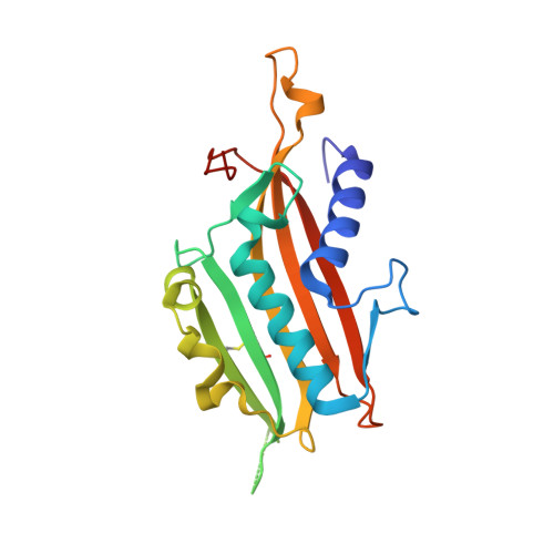
Structure of the Human TRPML2 Ion Channel Extracytosolic/Lumenal Domain.

Viet, K.K.Wagner, A.Schwickert, K.Hellwig, N.Brennich, M.Bader, N.Schirmeister, T.Morgner, N.Schindelin, H.Hellmich, U.A.

(2019) Structure 27: 1246-1257.e5

  • DOI: https://doi.org/10.1016/j.str.2019.04.016
  • Primary Citation of Related Structures:  
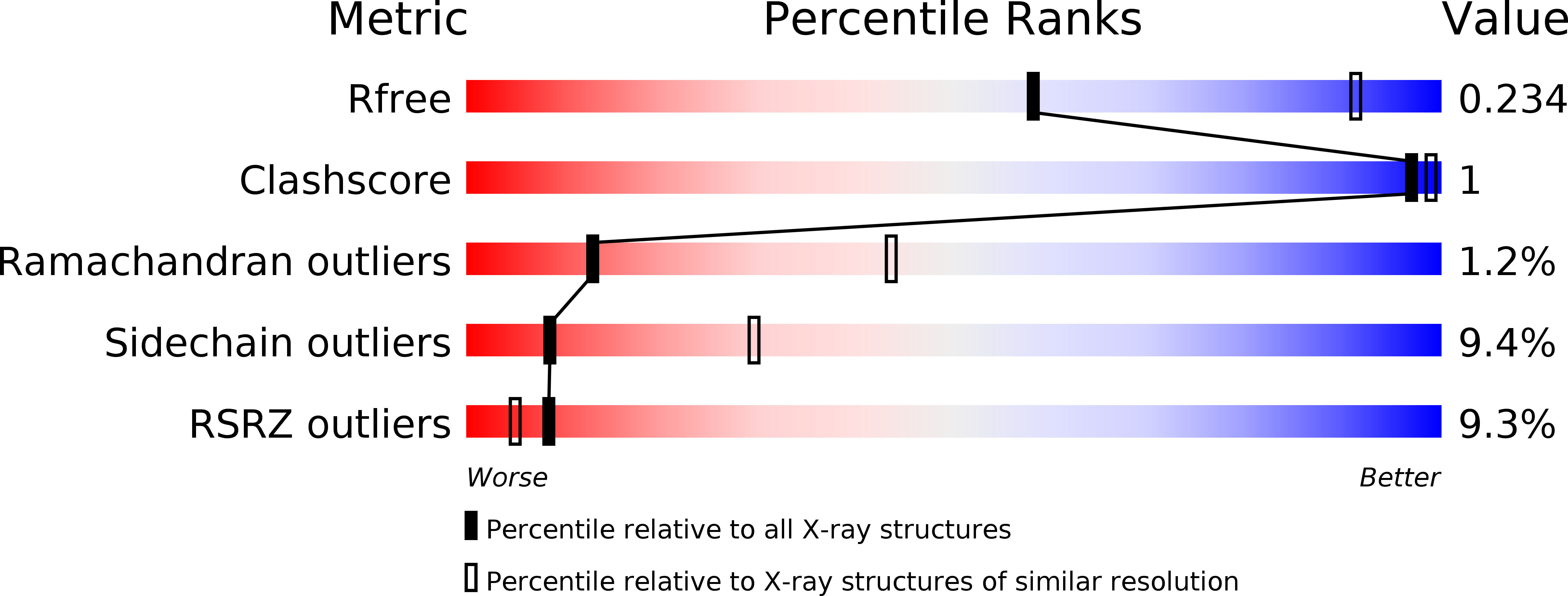
    6HRR, 6HRS

  • PubMed Abstract: 

    TRPML2 is the least structurally characterized mammalian transient receptor potential mucolipin ion channel. The TRPML family hallmark is a large extracytosolic/lumenal domain (ELD) between transmembrane helices S1 and S2. We present crystal structures of the tetrameric human TRPML2 ELD at pH 6.5 (2.0 Å) and 4.5 (2.95 Å), corresponding to the pH values in recycling endosomes and lysosomes. Isothermal titration calorimetry shows Ca 2+ binding to the highly acidic central pre-pore loop which is abrogated at low pH, in line with a pH-dependent channel regulation model. Small angle X-ray scattering confirms the ELD dimensions in solution. Changes in pH or Ca 2+ concentration do not affect the protein's secondary structure, but can influence ELD oligomer integrity according to native mass spectrometry. Our data thus complete the set of high-resolution views of human TRPML channel ELDs and reveal some structural responses to the conditions the TRPML2 ELD encounters as the channel traffics through the endolysosomal system.


  • Organizational Affiliation

    Institute for Pharmacy and Biochemistry, Johannes Gutenberg-Universität Mainz, 55128 Mainz, Germany; Center for Biomolecular Magnetic Resonance (BMRZ), Goethe-Universität, 60438 Frankfurt am Main, Germany.


Macromolecules
Find similar proteins by:  (by identity cutoff)  |  3D Structure
Entity ID: 1
MoleculeChains Sequence LengthOrganismDetailsImage
Mucolipin-2
A, B, C, D, E
A, B, C, D, E, F, G, H
199Homo sapiensMutation(s): 0 
Gene Names: MCOLN2
UniProt & NIH Common Fund Data Resources
Find proteins for Q8IZK6 (Homo sapiens)
Explore Q8IZK6 
Go to UniProtKB:  Q8IZK6
PHAROS:  Q8IZK6
GTEx:  ENSG00000153898 
Entity Groups  
Sequence Clusters30% Identity50% Identity70% Identity90% Identity95% Identity100% Identity
UniProt GroupQ8IZK6
Sequence Annotations
Expand
  • Reference Sequence
Experimental Data & Validation

Experimental Data

  • Method: X-RAY DIFFRACTION
  • Resolution: 2.95 Å
  • R-Value Free: 0.216 
  • R-Value Work: 0.182 
  • R-Value Observed: 0.184 
  • Space Group: P 1 21 1
Unit Cell:
Length ( Å )Angle ( ˚ )
a = 74.523α = 90
b = 142.473β = 104.49
c = 85.195γ = 90
Software Package:
Software NamePurpose
XDSdata reduction
Aimlessdata scaling
PHASERphasing
BUSTERrefinement
PDB_EXTRACTdata extraction

Structure Validation

View Full Validation Report


Entry History 

Deposition Data

Revision History  (Full details and data files)

  • Version 1.0: 2019-06-19
    Type: Initial release
  • Version 1.1: 2019-08-14
    Changes: Data collection, Database references
  • Version 1.2: 2024-01-24
    Changes: Data collection, Database references, Refinement description