6G1E

BEAT Fc with improved heterodimerization (Q3A-D84.4Q)


Experimental Data Snapshot

  • Method: X-RAY DIFFRACTION
  • Resolution: 1.88 Å
  • R-Value Free: 0.272 
  • R-Value Work: 0.231 
  • R-Value Observed: 0.233 

wwPDB Validation   3D Report Full Report


This is version 2.1 of the entry. See complete history


Literature

A single mutation increases heavy chain heterodimer assembly of bispecific antibodies by inducing structural disorder in one homodimer species.

Stutz, C.Blein, S.

(2020) J Biol Chem 

  • DOI: https://doi.org/10.1074/jbc.RA119.012335
  • Primary Citation of Related Structures:  
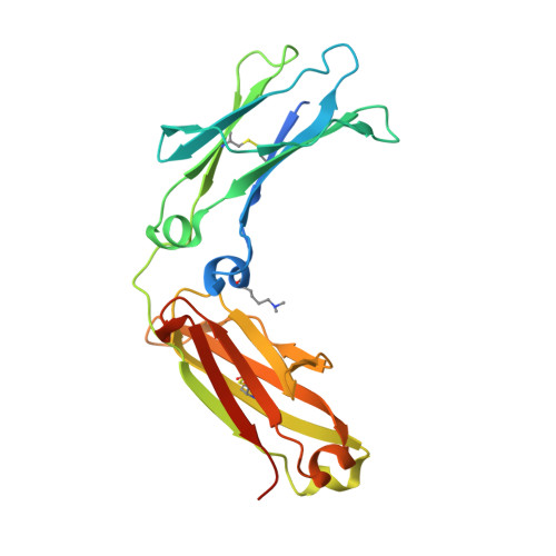
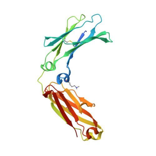
    6G1E

  • PubMed Abstract: 

    We previously reported efficient heavy-chain assembly of heterodimeric bispecific antibodies by exchanging the interdomain protein interface of the human IgG1 CH3 dimer with the protein interface of the constant α and β domains of the human T-cell receptor, a technology known as bispecific engagement by antibodies based on the T-cell receptor (BEAT). Efficient heterodimerization in mammalian cell transient transfections was observed, but levels were influenced by the nature of the binding arms, particularly in the Fab-scFv-Fc format. In this study, we report a single amino acid change that significantly and consistently improved the heterodimerization rate of this format (≥95%) by inducing partial disorder in one homodimer species without affecting the heterodimer. Correct folding and assembly of the heterodimer were confirmed by the high-resolution (1.88-1.98 Å) crystal structure presented here. Thermal stability and 1-anilinonaphthalene-8-sulfonic acid-binding experiments, comparing original BEAT, mutated BEAT, and "knobs-into-holes" interfaces, suggested a cooperative assembly process of heavy chains in heterodimers. The observed gain in stability of the interfaces could be classified in the following rank order: mutated BEAT > original BEAT > knobs-into-holes. We therefore propose that the superior cooperativity found in BEAT interfaces is the key driver of their greater performance. Furthermore, we show how the mutated BEAT interface can be exploited for the routine preparation of drug candidates, with minimal risk of homodimer contamination using a single Protein A chromatography step.


  • Organizational Affiliation

    Department of Antibody Engineering, Ichnos Sciences S.A., Biopôle Lausanne-Epalinges, Epalinges, Switzerland.


Macromolecules
Find similar proteins by:  (by identity cutoff)  |  3D Structure
Entity ID: 1
MoleculeChains Sequence LengthOrganismDetailsImage
Immunoglobulin heavy constant gamma 1,Immunoglobulin heavy constant gamma 3233Homo sapiensMutation(s): 11 
Gene Names: IGHG1IGHG3
UniProt & NIH Common Fund Data Resources
Find proteins for P01857 (Homo sapiens)
Explore P01857 
Go to UniProtKB:  P01857
PHAROS:  P01857
GTEx:  ENSG00000211896 
Find proteins for P01860 (Homo sapiens)
Explore P01860 
Go to UniProtKB:  P01860
PHAROS:  P01860
Entity Groups  
Sequence Clusters30% Identity50% Identity70% Identity90% Identity95% Identity100% Identity
UniProt GroupsP01857P01860
Sequence Annotations
Expand
  • Reference Sequence
Find similar proteins by:  (by identity cutoff)  |  3D Structure
Entity ID: 2
MoleculeChains Sequence LengthOrganismDetailsImage
Immunoglobulin gamma-1 heavy chain227Homo sapiensMutation(s): 16 
UniProt
Find proteins for P0DOX5 (Homo sapiens)
Explore P0DOX5 
Go to UniProtKB:  P0DOX5
Entity Groups  
Sequence Clusters30% Identity50% Identity70% Identity90% Identity95% Identity100% Identity
UniProt GroupP0DOX5
Sequence Annotations
Expand
  • Reference Sequence
Oligosaccharides

Help

Entity ID: 3
MoleculeChains Length2D Diagram Glycosylation3D Interactions
beta-D-galactopyranose-(1-4)-2-acetamido-2-deoxy-beta-D-glucopyranose-(1-2)-alpha-D-mannopyranose-(1-6)-[2-acetamido-2-deoxy-beta-D-glucopyranose-(1-2)-alpha-D-mannopyranose-(1-3)]beta-D-mannopyranose-(1-4)-2-acetamido-2-deoxy-beta-D-glucopyranose-(1-4)-[beta-L-fucopyranose-(1-6)]2-acetamido-2-deoxy-beta-D-glucopyranose
C, D
9N-Glycosylation
Glycosylation Resources
GlyTouCan:  G33244HE
GlyCosmos:  G33244HE
GlyGen:  G33244HE
Small Molecules
Modified Residues  1 Unique
IDChains TypeFormula2D DiagramParent
MLY
Query on MLY
A
L-PEPTIDE LINKINGC8 H18 N2 O2LYS
Experimental Data & Validation

Experimental Data

  • Method: X-RAY DIFFRACTION
  • Resolution: 1.88 Å
  • R-Value Free: 0.272 
  • R-Value Work: 0.231 
  • R-Value Observed: 0.233 
  • Space Group: P 21 21 21
Unit Cell:
Length ( Å )Angle ( ˚ )
a = 49.65α = 90
b = 73.02β = 90
c = 140.93γ = 90
Software Package:
Software NamePurpose
REFMACrefinement
MOSFLMdata reduction
SCALAdata scaling
PHASERphasing

Structure Validation

View Full Validation Report


Entry History 

Deposition Data

Revision History  (Full details and data files)

  • Version 1.0: 2019-04-10
    Type: Initial release
  • Version 1.1: 2020-06-03
    Changes: Data collection, Database references
  • Version 2.0: 2020-07-29
    Type: Remediation
    Reason: Carbohydrate remediation
    Changes: Advisory, Atomic model, Data collection, Derived calculations, Structure summary
  • Version 2.1: 2024-01-17
    Changes: Data collection, Database references, Derived calculations, Refinement description, Structure summary