Solution NMR investigation on the structure and function of the isolated J-domain from Sis1: Evidence of transient inter-domain interactions in the full-length protein.
Pinheiro, G.M.S., Amorim, G.C., Iqbal, A., Almeida, F.C.L., Ramos, C.H.I.(2019) Arch Biochem Biophys 669: 71-79
- PubMed: 31141701
- DOI: https://doi.org/10.1016/j.abb.2019.05.020
- Primary Citation of Related Structures:
6D6X - PubMed Abstract:
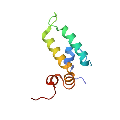
J-domain/Hsp40 proteins cooperate in aiding with folding in the cell by binding partially folded client proteins and delivering them to be folded by Hsp70. The delivery occurs concomitantly to the stimulation of the ATPase activity of Hsp70 via the N-terminally located J-domain. Although several lines of investigation have been used to study J-domain proteins, the presence of highly flexible domains (G/F- and G/M-rich) hold up obtaining a detailed full-length structure. In this work, we present the high-resolution structure of the J-domain and the N-terminal part of the G/F domain of Sis1, solved by NMR, and used chemical-shift perturbation approaches to further study the structure/function relationship of the Sis1/Hsp70 interaction. When the J-domain was compared to the full-length protein and to a G/M domain deletion mutant, an internal interaction patch formed by hydrophobic and positively charged residues (V2, D9, R27, T39, F52 and R73) was identified. Curiously, the same patch is protected by internal contacts in the full-length protein and, in combination with the loop containing the conserved HPD motif, participates in the interaction with Hsp70. Combined, these results suggest that the J-domain in the full-length Sis1 is in a transient intermediate conformation, in which its interacting patch is protected and, at the same time, also in a favorable condition to bind Hsp70, facilitating the interaction between the two proteins. Finally, 1D NMR experiments showed that the addition of ATP is followed by the disruption of the J-domain/Hsp70 complex, a necessary step for aiding the folding of the client protein.
- Institute of Chemistry, University of Campinas UNICAMP, Campinas, SP, Brazil.
Organizational Affiliation: