6C5F

Human D-Dopachrome tautomerase (D-DT)/ macrophage migration inhibitory factor 2 (MIF2) complexed with the selective inhibitor 4-CPPC


Experimental Data Snapshot

  • Method: X-RAY DIFFRACTION
  • Resolution: 1.40 Å
  • R-Value Free: 0.157 
  • R-Value Work: 0.118 
  • R-Value Observed: 0.119 

wwPDB Validation   3D Report Full Report


Ligand Structure Quality Assessment 


This is version 1.2 of the entry. See complete history


Literature

Structural Plasticity in the C-Terminal Region of Macrophage Migration Inhibitory Factor-2 Is Associated with an Induced Fit Mechanism for a Selective Inhibitor.

Pantouris, G.Bucala, R.Lolis, E.J.

(2018) Biochemistry 57: 3599-3605

  • DOI: https://doi.org/10.1021/acs.biochem.8b00344
  • Primary Citation of Related Structures:  
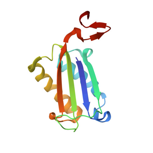
    6C5F

  • PubMed Abstract: 

    We report the first reversible and selective small molecule inhibitor of pro-inflammatory protein macrophage migration inhibitory factor-2 (also known as MIF-2 or d-DT). 4-(3-Carboxyphenyl)-2,5-pyridinedicarboxylic acid (4-CPPC) shows competitive binding with a 13-fold selectivity for human MIF-2 versus human MIF-1. The crystal structure of MIF-2 complexed with 4-CPPC reveals an induced fit mechanism that is not observed in the numerous MIF-1/inhibitor complexes. Crystallographic analysis demonstrates the structural source of 4-CPPC binding and selectivity for MIF-2. 4-CPPC can be employed to reveal previously unrecognized functions of MIF-1 in biological systems in which both MIF-1 and MIF-2 are expressed, to improve our knowledge of the MIF family of proteins, and to provide new mechanistic insights that can be utilized for the development of potent and selective pharmacological modulators of MIF-2.


  • Organizational Affiliation

    Department of Pharmacology , Yale School of Medicine , New Haven , Connecticut 06510 , United States.


Macromolecules
Find similar proteins by:  (by identity cutoff)  |  3D Structure
Entity ID: 1
MoleculeChains Sequence LengthOrganismDetailsImage
D-dopachrome decarboxylase
A, B, C
117Homo sapiensMutation(s): 0 
Gene Names: DDT
EC: 4.1.1.84
UniProt & NIH Common Fund Data Resources
Find proteins for P30046 (Homo sapiens)
Explore P30046 
Go to UniProtKB:  P30046
PHAROS:  P30046
GTEx:  ENSG00000099977 
Entity Groups  
Sequence Clusters30% Identity50% Identity70% Identity90% Identity95% Identity100% Identity
UniProt GroupP30046
Sequence Annotations
Expand
  • Reference Sequence
Small Molecules
Ligands 1 Unique
IDChains Name / Formula / InChI Key2D Diagram3D Interactions
7L9
Query on 7L9

Download Ideal Coordinates CCD File 
D [auth B]4-(3-carboxyphenyl)pyridine-2,5-dicarboxylic acid
C14 H9 N O6
NBVHLJGGHJLTGM-UHFFFAOYSA-N
Binding Affinity Annotations 
IDSourceBinding Affinity
7L9 Binding MOAD:  6C5F Ki: 3.30e+4 (nM) from 1 assay(s)
Experimental Data & Validation

Experimental Data

  • Method: X-RAY DIFFRACTION
  • Resolution: 1.40 Å
  • R-Value Free: 0.157 
  • R-Value Work: 0.118 
  • R-Value Observed: 0.119 
  • Space Group: P 3
Unit Cell:
Length ( Å )Angle ( ˚ )
a = 83.936α = 90
b = 83.936β = 90
c = 40.479γ = 120
Software Package:
Software NamePurpose
HKL-2000data scaling
MOLREPphasing
REFMACrefinement
PDB_EXTRACTdata extraction
HKL-2000data reduction

Structure Validation

View Full Validation Report


Ligand Structure Quality Assessment 


Entry History 

Deposition Data

Revision History  (Full details and data files)

  • Version 1.0: 2018-06-13
    Type: Initial release
  • Version 1.1: 2018-07-11
    Changes: Data collection, Database references
  • Version 1.2: 2023-10-04
    Changes: Data collection, Database references, Refinement description