6BST

CRYSTAL STRUCTURE OF Z-DNA WITH UNTYPICALLY COORDINATED CA2+ ION


Experimental Data Snapshot

  • Method: X-RAY DIFFRACTION
  • Resolution: 1.45 Å
  • R-Value Free: 0.172 
  • R-Value Work: 0.121 
  • R-Value Observed: 0.121 

wwPDB Validation   3D Report Full Report


This is version 1.3 of the entry. See complete history


Literature

The crystal structure of Z-DNA with untypically coordinated Ca

Luo, Z.Dauter, Z.

(2018) J Biol Inorg Chem 23: 253-259

  • DOI: https://doi.org/10.1007/s00775-017-1526-4
  • Primary Citation of Related Structures:  
    6BST

  • PubMed Abstract: 

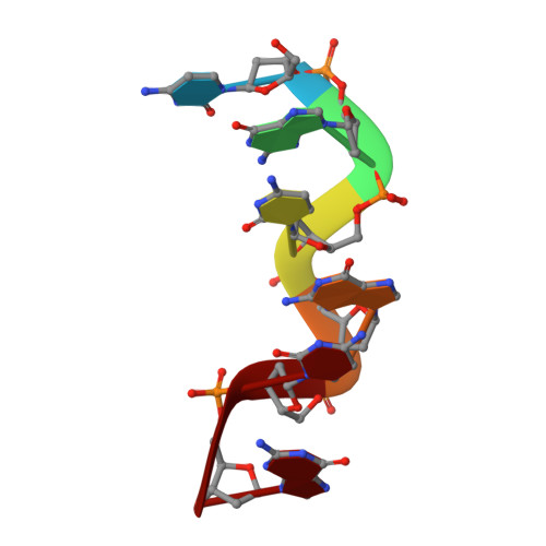
    DNA oligomer duplexes with alternating cytosines and guanines in their sequence tend to form helices of the Z-DNA type, where the sugar and phosphate backbone forms a left-handed helix in a zigzag fashion with a repeat of two successive Watson-Crick pairs of nucleotides. Z-DNA duplexes often crystallize in complexes with diverse metal ions interacting with polar DNA atoms in various ways. This work describes the high-resolution crystal structure of a Z-DNA d(CGCGCG) 2 duplex in complex with Ca 2+ ions, unusually coordinated as an approximate pentagonal bipyramid by two neighboring guanines through their O6 and N7 atoms and a water molecule in the equatorial plane and a phosphate oxygen atom and another water molecule in the apical positions.


  • Organizational Affiliation

    Synchrotron Radiation Research Section, MCL, National Cancer Institute, Argonne National Laboratory, Argonne, IL, 60439, USA.


Macromolecules

Find similar nucleic acids by:  Sequence   |   3D Structure  

Entity ID: 1
MoleculeChains LengthOrganismImage
DNA (5'-D(*CP*GP*CP*GP*CP*G)-3')
A, B
6synthetic construct
Sequence Annotations
Expand
  • Reference Sequence
Small Molecules
Ligands 2 Unique
IDChains Name / Formula / InChI Key2D Diagram3D Interactions
SR0
Query on SR0

Download Ideal Coordinates CCD File 
C [auth A]N~1~-(3-azaniumylpropyl)butane-1,4-diaminium
C7 H22 N3
ATHGHQPFGPMSJY-UHFFFAOYSA-Q
CA
Query on CA

Download Ideal Coordinates CCD File 
D [auth B]CALCIUM ION
Ca
BHPQYMZQTOCNFJ-UHFFFAOYSA-N
Experimental Data & Validation

Experimental Data

  • Method: X-RAY DIFFRACTION
  • Resolution: 1.45 Å
  • R-Value Free: 0.172 
  • R-Value Work: 0.121 
  • R-Value Observed: 0.121 
  • Space Group: P 21 21 21
Unit Cell:
Length ( Å )Angle ( ˚ )
a = 17.76α = 90
b = 28.82β = 90
c = 42.36γ = 90
Software Package:
Software NamePurpose
REFMACrefinement
HKL-2000data reduction
HKL-2000data scaling
PHASERphasing

Structure Validation

View Full Validation Report


Entry History 

Deposition Data

Revision History  (Full details and data files)

  • Version 1.0: 2017-12-20
    Type: Initial release
  • Version 1.1: 2018-01-10
    Changes: Database references
  • Version 1.2: 2018-02-28
    Changes: Database references
  • Version 1.3: 2023-10-04
    Changes: Data collection, Database references, Derived calculations, Refinement description