6AKD

Crystal structure of IdnL7


Experimental Data Snapshot

  • Method: X-RAY DIFFRACTION
  • Resolution: 2.10 Å
  • R-Value Free: 0.220 
  • R-Value Work: 0.177 
  • R-Value Observed: 0.179 

wwPDB Validation   3D Report Full Report


Ligand Structure Quality Assessment 


This is version 1.2 of the entry. See complete history


Literature

Functional and structural characterization of IdnL7, an adenylation enzyme involved in incednine biosynthesis.

Cieslak, J.Miyanaga, A.Takaishi, M.Kudo, F.Eguchi, T.

(2019) Acta Crystallogr F Struct Biol Commun 75: 299-306

  • DOI: https://doi.org/10.1107/S2053230X19002863
  • Primary Citation of Related Structures:  
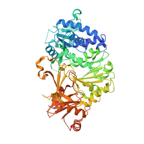
    6AKD

  • PubMed Abstract: 

    Adenylation enzymes play an important role in the selective incorporation of the cognate carboxylate substrates in natural product biosynthesis. Here, the biochemical and structural characterization of the adenylation enzyme IdnL7, which is involved in the biosynthesis of the macrolactam polyketide antibiotic incednine, is reported. Biochemical analysis showed that IdnL7 selects and activates several small amino acids. The structure of IdnL7 in complex with an L-alanyl-adenylate intermediate mimic, 5'-O-[N-(L-alanyl)sulfamoyl]adenosine, was determined at 2.1 Å resolution. The structure of IdnL7 explains the broad substrate specificity of IdnL7 towards small L-amino acids.


  • Organizational Affiliation

    Department of Chemistry and Materials Science, Tokyo Institute of Technology, 2-12-1 O-okayama, Meguro-ku, Tokyo 152-8551, Japan.


Macromolecules
Find similar proteins by:  (by identity cutoff)  |  3D Structure
Entity ID: 1
MoleculeChains Sequence LengthOrganismDetailsImage
AMP-dependent synthetase and ligase538Streptomyces sp. ML694-90F3Mutation(s): 0 
Gene Names: idnL7
UniProt
Find proteins for A0A077KUW8 (Streptomyces sp. ML694-90F3)
Explore A0A077KUW8 
Go to UniProtKB:  A0A077KUW8
Entity Groups  
Sequence Clusters30% Identity50% Identity70% Identity90% Identity95% Identity100% Identity
UniProt GroupA0A077KUW8
Sequence Annotations
Expand
  • Reference Sequence
Experimental Data & Validation

Experimental Data

  • Method: X-RAY DIFFRACTION
  • Resolution: 2.10 Å
  • R-Value Free: 0.220 
  • R-Value Work: 0.177 
  • R-Value Observed: 0.179 
  • Space Group: P 41 21 2
Unit Cell:
Length ( Å )Angle ( ˚ )
a = 68.526α = 90
b = 68.526β = 90
c = 225.169γ = 90
Software Package:
Software NamePurpose
REFMACrefinement
HKL-2000data reduction
HKL-2000data scaling
MOLREPphasing

Structure Validation

View Full Validation Report


Ligand Structure Quality Assessment 


Entry History & Funding Information

Deposition Data


Funding OrganizationLocationGrant Number
Japan Society for the Promotion of ScienceJapan25850050

Revision History  (Full details and data files)

  • Version 1.0: 2019-03-06
    Type: Initial release
  • Version 1.1: 2019-04-17
    Changes: Data collection, Database references
  • Version 1.2: 2023-11-22
    Changes: Data collection, Database references, Refinement description