6A2U

Crystal structure of gamma-alpha subunit complex from Burkholderia cepacia FAD glucose dehydrogenase


Experimental Data Snapshot

  • Method: X-RAY DIFFRACTION
  • Resolution: 2.60 Å
  • R-Value Free: 0.261 
  • R-Value Work: 0.204 
  • R-Value Observed: 0.207 

wwPDB Validation   3D Report Full Report


Ligand Structure Quality Assessment 


This is version 1.3 of the entry. See complete history


Literature

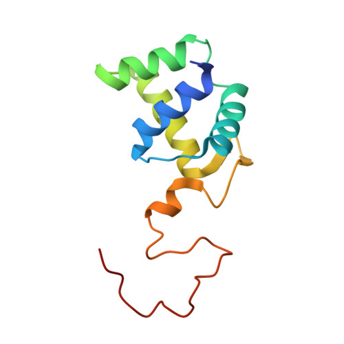
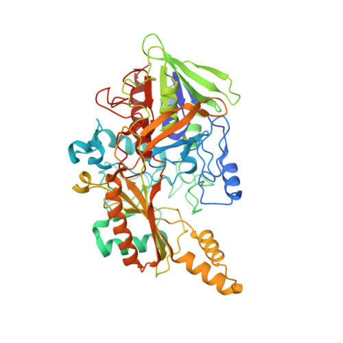
X-ray structure of the direct electron transfer-type FAD glucose dehydrogenase catalytic subunit complexed with a hitchhiker protein.

Yoshida, H.Kojima, K.Shiota, M.Yoshimatsu, K.Yamazaki, T.Ferri, S.Tsugawa, W.Kamitori, S.Sode, K.

(2019) Acta Crystallogr D Struct Biol 75: 841-851

  • DOI: https://doi.org/10.1107/S2059798319010878
  • Primary Citation of Related Structures:  
    6A2U

  • PubMed Abstract: 

    The bacterial flavin adenine dinucleotide (FAD)-dependent glucose dehydrogenase complex derived from Burkholderia cepacia (BcGDH) is a representative molecule of direct electron transfer-type FAD-dependent dehydrogenase complexes. In this study, the X-ray structure of BcGDHγα, the catalytic subunit (α-subunit) of BcGDH complexed with a hitchhiker protein (γ-subunit), was determined. The most prominent feature of this enzyme is the presence of the 3Fe-4S cluster, which is located at the surface of the catalytic subunit and functions in intramolecular and intermolecular electron transfer from FAD to the electron-transfer subunit. The structure of the complex revealed that these two molecules are connected through disulfide bonds and hydrophobic interactions, and that the formation of disulfide bonds is required to stabilize the catalytic subunit. The structure of the complex revealed the putative position of the electron-transfer subunit. A comparison of the structures of BcGDHγα and membrane-bound fumarate reductases suggested that the whole BcGDH complex, which also includes the membrane-bound β-subunit containing three heme c moieties, may form a similar overall structure to fumarate reductases, thus accomplishing effective electron transfer.


  • Organizational Affiliation

    Life Science Research Center and Faculty of Medicine, Kagawa University, 1750-1 Ikenobe, Miki-cho, Kita-gun, Kagawa 761-0793, Japan.


Macromolecules
Find similar proteins by:  (by identity cutoff)  |  3D Structure
Entity ID: 1
MoleculeChains Sequence LengthOrganismDetailsImage
Twin-arginine translocation pathway signal
A, C
121Burkholderia cepaciaMutation(s): 0 
UniProt
Find proteins for A0A0H3KLY3 (Burkholderia multivorans (strain ATCC 17616 / 249))
Explore A0A0H3KLY3 
Go to UniProtKB:  A0A0H3KLY3
Entity Groups  
Sequence Clusters30% Identity50% Identity70% Identity90% Identity95% Identity100% Identity
UniProt GroupA0A0H3KLY3
Sequence Annotations
Expand
  • Reference Sequence
Find similar proteins by:  (by identity cutoff)  |  3D Structure
Entity ID: 2
MoleculeChains Sequence LengthOrganismDetailsImage
Glucose dehydrogenase
B, D
545Burkholderia cepaciaMutation(s): 0 
Gene Names: gdhAlpha
EC: 1.1.5.9
UniProt
Find proteins for Q8GQE7 (Burkholderia cepacia)
Explore Q8GQE7 
Go to UniProtKB:  Q8GQE7
Entity Groups  
Sequence Clusters30% Identity50% Identity70% Identity90% Identity95% Identity100% Identity
UniProt GroupQ8GQE7
Sequence Annotations
Expand
  • Reference Sequence
Experimental Data & Validation

Experimental Data

  • Method: X-RAY DIFFRACTION
  • Resolution: 2.60 Å
  • R-Value Free: 0.261 
  • R-Value Work: 0.204 
  • R-Value Observed: 0.207 
  • Space Group: P 65 2 2
Unit Cell:
Length ( Å )Angle ( ˚ )
a = 110.521α = 90
b = 110.521β = 90
c = 524.877γ = 120
Software Package:
Software NamePurpose
REFMACrefinement
SCALEPACKdata scaling
PDB_EXTRACTdata extraction
HKL-2000data reduction
MOLREPphasing

Structure Validation

View Full Validation Report


Ligand Structure Quality Assessment 


Entry History 

Deposition Data

Revision History  (Full details and data files)

  • Version 1.0: 2019-06-19
    Type: Initial release
  • Version 1.1: 2019-09-04
    Changes: Data collection, Database references
  • Version 1.2: 2019-09-18
    Changes: Data collection, Database references
  • Version 1.3: 2024-04-03
    Changes: Data collection, Database references, Derived calculations, Refinement description