6SM1

Wild type immunoglobulin light chain (WT-1)


Experimental Data Snapshot

  • Method: X-RAY DIFFRACTION
  • Resolution: 1.55 Å
  • R-Value Free: 0.165 
  • R-Value Work: 0.142 
  • R-Value Observed: 0.143 

wwPDB Validation   3D Report Full Report


This is version 1.1 of the entry. See complete history


Literature

Fatal amyloid formation in a patient's antibody light chain is caused by a single point mutation.

Kazman, P.Vielberg, M.T.Pulido Cendales, M.D.Hunziger, L.Weber, B.Hegenbart, U.Zacharias, M.Kohler, R.Schonland, S.Groll, M.Buchner, J.

(2020) Elife 9

  • DOI: https://doi.org/10.7554/eLife.52300
  • Primary Citation of Related Structures:  
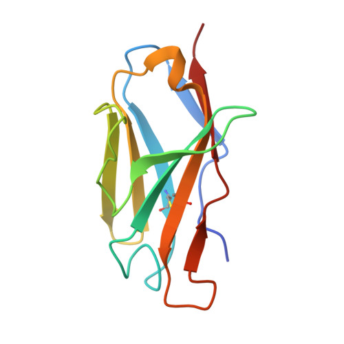
    6SM1, 6SM2

  • PubMed Abstract: 

    In systemic light chain amyloidosis, an overexpressed antibody light chain (LC) forms fibrils which deposit in organs and cause their failure. While it is well-established that mutations in the LC's V L domain are important prerequisites, the mechanisms which render a patient LC amyloidogenic are ill-defined. In this study, we performed an in-depth analysis of the factors and mutations responsible for the pathogenic transformation of a patient-derived λ LC, by recombinantly expressing variants in E. coli . We show that proteolytic cleavage of the patient LC resulting in an isolated V L domain is essential for fibril formation. Out of 11 mutations in the patient V L , only one, a leucine to valine mutation, is responsible for fibril formation. It disrupts a hydrophobic network rendering the C-terminal segment of V L more dynamic and decreasing domain stability. Thus, the combination of proteolytic cleavage and the destabilizing mutation trigger conformational changes that turn the LC pathogenic.


  • Organizational Affiliation

    Center for Integrated Protein Science Munich at the Department Chemie, Technische Universität München, Garching, Germany.


Macromolecules
Find similar proteins by:  (by identity cutoff)  |  3D Structure
Entity ID: 1
MoleculeChains Sequence LengthOrganismDetailsImage
Immunoglobulin lambda variable 2-14114Homo sapiensMutation(s): 0 
Gene Names: IGLV2-14
UniProt & NIH Common Fund Data Resources
Find proteins for P01704 (Homo sapiens)
Explore P01704 
Go to UniProtKB:  P01704
GTEx:  ENSG00000211666 
Entity Groups  
Sequence Clusters30% Identity50% Identity70% Identity90% Identity95% Identity100% Identity
UniProt GroupP01704
Sequence Annotations
Expand
  • Reference Sequence
Experimental Data & Validation

Experimental Data

  • Method: X-RAY DIFFRACTION
  • Resolution: 1.55 Å
  • R-Value Free: 0.165 
  • R-Value Work: 0.142 
  • R-Value Observed: 0.143 
  • Space Group: P 63 2 2
Unit Cell:
Length ( Å )Angle ( ˚ )
a = 104.5α = 90
b = 104.5β = 90
c = 55.22γ = 120
Software Package:
Software NamePurpose
REFMACrefinement
PDB_EXTRACTdata extraction
XDSdata reduction
XSCALEdata scaling
PHASERphasing

Structure Validation

View Full Validation Report


Entry History & Funding Information

Deposition Data


Funding OrganizationLocationGrant Number
German Research FoundationGermanySFB1035

Revision History  (Full details and data files)

  • Version 1.0: 2020-03-18
    Type: Initial release
  • Version 1.1: 2024-01-24
    Changes: Data collection, Database references, Derived calculations, Refinement description