6EHO

Dimer of the Sortilin Vps10p domain at low pH


Experimental Data Snapshot

  • Method: X-RAY DIFFRACTION
  • Resolution: 3.50 Å
  • R-Value Free: 0.272 
  • R-Value Work: 0.232 
  • R-Value Observed: 0.233 

wwPDB Validation   3D Report Full Report


This is version 2.1 of the entry. See complete history


Literature

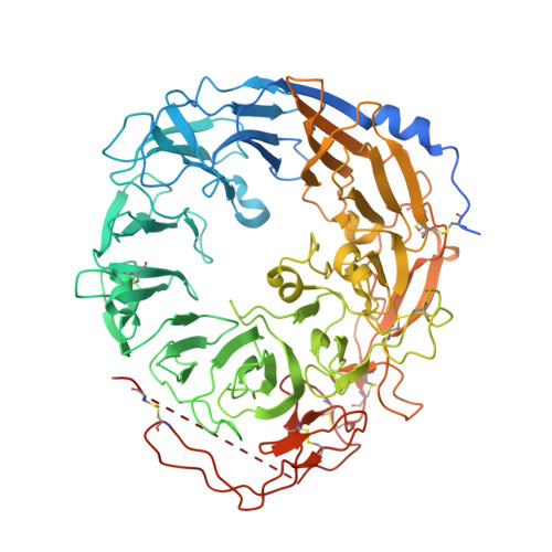
Acidic Environment Induces Dimerization and Ligand Binding Site Collapse in the Vps10p Domain of Sortilin.

Januliene, D.Andersen, J.L.Nielsen, J.A.Quistgaard, E.M.Hansen, M.Strandbygaard, D.Moeller, A.Petersen, C.M.Madsen, P.Thirup, S.S.

(2017) Structure 25: 1809-1819.e3

  • DOI: https://doi.org/10.1016/j.str.2017.09.015
  • Primary Citation of Related Structures:  
    6EHO

  • PubMed Abstract: 

    Sortilin is a neuronal receptor involved in transmembrane signaling, endocytosis, and intracellular sorting of proteins. It cycles through a number of cellular compartments where it encounters various acidic conditions. The crystal structure of the sortilin ectodomain has previously been determined at neutral pH. Here, we present the 3.5-Å resolution crystal structure of sortilin at pH 5.5, which represents an environment similar to that of late endosomes, where ligands are released. The structure reveals an overall distortion of the 10-bladed β-propeller domain. This distortion and specific conformational changes, caused by protonation of a number of histidine residues, render the currently known binding sites unavailable for ligand binding. Access to the binding sites is furthermore blocked by a reversible and pH-dependent formation of tight sortilin dimers, also confirmed by electron microscopy, size-exclusion chromatography, and mutational studies. This study reveals how sortilin binding sites are disrupted and explains pH-dependent ligand affinity.


  • Organizational Affiliation

    MIND Centre, Department of Molecular Biology and Genetics, Aarhus University, 8000 Aarhus, Denmark; Department of Structural Biology, Max Planck Institute of Biophysics, 60438 Frankfurt am Main, Germany.


Macromolecules
Find similar proteins by:  (by identity cutoff)  |  3D Structure
Entity ID: 1
MoleculeChains Sequence LengthOrganismDetailsImage
Sortilin723Homo sapiensMutation(s): 2 
Gene Names: SORT1
UniProt & NIH Common Fund Data Resources
Find proteins for Q99523 (Homo sapiens)
Explore Q99523 
Go to UniProtKB:  Q99523
PHAROS:  Q99523
GTEx:  ENSG00000134243 
Entity Groups  
Sequence Clusters30% Identity50% Identity70% Identity90% Identity95% Identity100% Identity
UniProt GroupQ99523
Sequence Annotations
Expand
  • Reference Sequence
Oligosaccharides

Help

Entity ID: 2
MoleculeChains Length2D Diagram Glycosylation3D Interactions
alpha-D-mannopyranose-(1-3)-[alpha-D-mannopyranose-(1-6)]beta-D-mannopyranose-(1-4)-2-acetamido-2-deoxy-beta-D-glucopyranose-(1-4)-2-acetamido-2-deoxy-beta-D-glucopyranose
B
5N-Glycosylation
Glycosylation Resources
GlyTouCan:  G22768VO
GlyCosmos:  G22768VO
GlyGen:  G22768VO
Entity ID: 3
MoleculeChains Length2D Diagram Glycosylation3D Interactions
beta-D-mannopyranose-(1-4)-2-acetamido-2-deoxy-beta-D-glucopyranose-(1-4)-2-acetamido-2-deoxy-beta-D-glucopyranose
C
3N-Glycosylation
Glycosylation Resources
GlyTouCan:  G15407YE
GlyCosmos:  G15407YE
GlyGen:  G15407YE
Entity ID: 4
MoleculeChains Length2D Diagram Glycosylation3D Interactions
2-acetamido-2-deoxy-beta-D-glucopyranose-(1-4)-2-acetamido-2-deoxy-beta-D-glucopyranose
D
2N-Glycosylation
Glycosylation Resources
GlyTouCan:  G42666HT
GlyCosmos:  G42666HT
GlyGen:  G42666HT
Experimental Data & Validation

Experimental Data

  • Method: X-RAY DIFFRACTION
  • Resolution: 3.50 Å
  • R-Value Free: 0.272 
  • R-Value Work: 0.232 
  • R-Value Observed: 0.233 
  • Space Group: I 21 3
Unit Cell:
Length ( Å )Angle ( ˚ )
a = 189.01α = 90
b = 189.01β = 90
c = 189.01γ = 90
Software Package:
Software NamePurpose
PHENIXrefinement
XDSdata reduction
XSCALEdata scaling
PHASERphasing

Structure Validation

View Full Validation Report


Entry History & Funding Information

Deposition Data


Funding OrganizationLocationGrant Number
Lundbeck FoundationDenmark--

Revision History  (Full details and data files)

  • Version 1.0: 2017-12-06
    Type: Initial release
  • Version 1.1: 2017-12-13
    Changes: Database references
  • Version 2.0: 2020-07-29
    Type: Remediation
    Reason: Carbohydrate remediation
    Changes: Atomic model, Data collection, Derived calculations, Structure summary
  • Version 2.1: 2024-01-17
    Changes: Data collection, Database references, Derived calculations, Refinement description, Structure summary