De novo design of self-assembling helical protein filaments.
Shen, H., Fallas, J.A., Lynch, E., Sheffler, W., Parry, B., Jannetty, N., Decarreau, J., Wagenbach, M., Vicente, J.J., Chen, J., Wang, L., Dowling, Q., Oberdorfer, G., Stewart, L., Wordeman, L., De Yoreo, J., Jacobs-Wagner, C., Kollman, J., Baker, D.(2018) Science 362: 705-709
- PubMed: 30409885
- DOI: https://doi.org/10.1126/science.aau3775
- Primary Citation of Related Structures:
6E9R, 6E9T, 6E9V, 6E9X, 6E9Y, 6E9Z - PubMed Abstract:
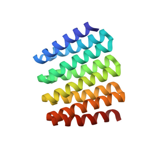
We describe a general computational approach to designing self-assembling helical filaments from monomeric proteins and use this approach to design proteins that assemble into micrometer-scale filaments with a wide range of geometries in vivo and in vitro. Cryo-electron microscopy structures of six designs are close to the computational design models. The filament building blocks are idealized repeat proteins, and thus the diameter of the filaments can be systematically tuned by varying the number of repeat units. The assembly and disassembly of the filaments can be controlled by engineered anchor and capping units built from monomers lacking one of the interaction surfaces. The ability to generate dynamic, highly ordered structures that span micrometers from protein monomers opens up possibilities for the fabrication of new multiscale metamaterials.
Organizational Affiliation:
Institute for Protein Design, University of Washington, Seattle, WA 98195, USA.