


Crystal Structure of Bifunctional protein FolD from Helicobacter pylori
SSGCID, Delker, S.L., Dranow, D.M., Lorimer, D., Edwards, T.E.To be published.
Experimental Data Snapshot
Starting Model: experimental
View more details
wwPDB Validation 3D Report Full Report

Entity ID: 1 | |||||
|---|---|---|---|---|---|
| Molecule | Chains | Sequence Length | Organism | Details | Image |
| Bifunctional protein FolD | 298 | Helicobacter pylori G27 | Mutation(s): 0 Gene Names: folD, HPG27_536 EC: 1.5.1.5 (PDB Primary Data), 3.5.4.9 (PDB Primary Data) | 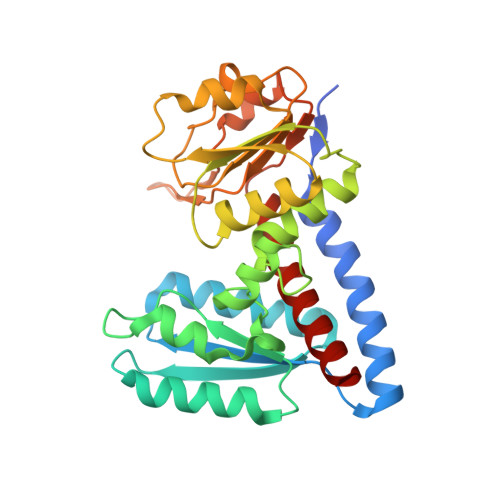 | |
UniProt | |||||
Find proteins for B5Z6U8 (Helicobacter pylori (strain G27)) Explore B5Z6U8 Go to UniProtKB: B5Z6U8 | |||||
Entity Groups | |||||
| Sequence Clusters | 30% Identity50% Identity70% Identity90% Identity95% Identity100% Identity | ||||
| UniProt Group | B5Z6U8 | ||||
Sequence AnnotationsExpand | |||||
| |||||
| Ligands 2 Unique | |||||
|---|---|---|---|---|---|
| ID | Chains | Name / Formula / InChI Key | 2D Diagram | 3D Interactions | |
| GOL Query on GOL | D [auth A], E [auth A], F [auth A] | GLYCEROL C3 H8 O3 PEDCQBHIVMGVHV-UHFFFAOYSA-N |  | ||
| NA Query on NA | B [auth A], C [auth A] | SODIUM ION Na FKNQFGJONOIPTF-UHFFFAOYSA-N |  | ||
| Length ( Å ) | Angle ( ˚ ) |
|---|---|
| a = 69.88 | α = 90 |
| b = 109.04 | β = 90 |
| c = 83.8 | γ = 90 |
| Software Name | Purpose |
|---|---|
| XSCALE | data scaling |
| PHENIX | refinement |
| PDB_EXTRACT | data extraction |
| MOLREP | phasing |


