


Crystal structure of PI3K complex with an inhibitor
Song, K., Yang, X., Zhao, Y., Jian, Z.To be published.
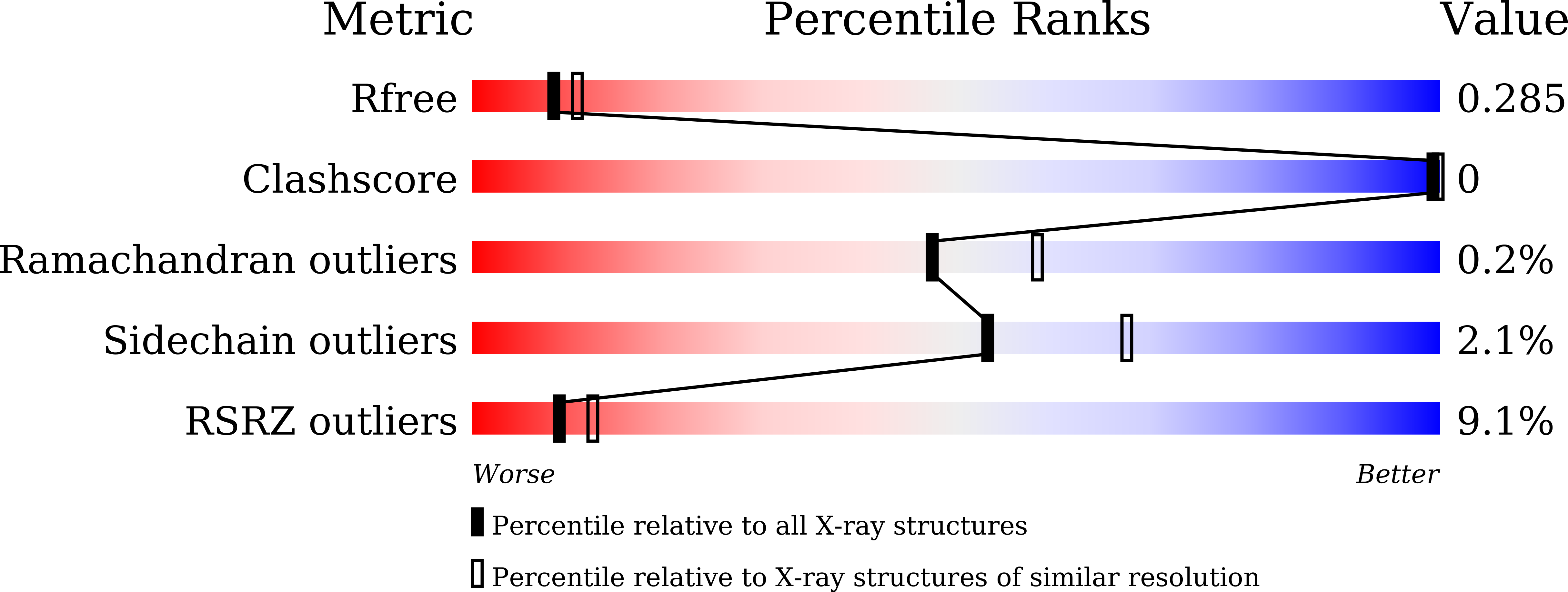
Experimental Data Snapshot
Entity ID: 1 | |||||
|---|---|---|---|---|---|
| Molecule | Chains | Sequence Length | Organism | Details | Image |
| Phosphatidylinositol 4,5-bisphosphate 3-kinase catalytic subunit alpha isoform | 1,052 | Homo sapiens | Mutation(s): 0 Gene Names: PIK3CA EC: 2.7.1.153 (PDB Primary Data), 2.7.11.1 (PDB Primary Data) | 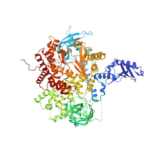 | |
UniProt & NIH Common Fund Data Resources | |||||
Find proteins for P42336 (Homo sapiens) Explore P42336 Go to UniProtKB: P42336 | |||||
PHAROS: P42336 GTEx: ENSG00000121879 | |||||
Entity Groups | |||||
| Sequence Clusters | 30% Identity50% Identity70% Identity90% Identity95% Identity100% Identity | ||||
| UniProt Group | P42336 | ||||
Sequence AnnotationsExpand | |||||
| |||||
Entity ID: 2 | |||||
|---|---|---|---|---|---|
| Molecule | Chains | Sequence Length | Organism | Details | Image |
| Phosphatidylinositol 3-kinase regulatory subunit alpha | 277 | Homo sapiens | Mutation(s): 0 Gene Names: PIK3R1, GRB1 |  | |
UniProt & NIH Common Fund Data Resources | |||||
Find proteins for P27986 (Homo sapiens) Explore P27986 Go to UniProtKB: P27986 | |||||
PHAROS: P27986 GTEx: ENSG00000145675 | |||||
Entity Groups | |||||
| Sequence Clusters | 30% Identity50% Identity70% Identity90% Identity95% Identity100% Identity | ||||
| UniProt Group | P27986 | ||||
Sequence AnnotationsExpand | |||||
| |||||
| Ligands 4 Unique | |||||
|---|---|---|---|---|---|
| ID | Chains | Name / Formula / InChI Key | 2D Diagram | 3D Interactions | |
| 84R Query on 84R | C [auth A] | 3-azanyl-5-(4-morpholin-4-ylthieno[3,2-d]pyrimidin-2-yl)phenol C16 H16 N4 O2 S GTKGIORNQYOWBR-UHFFFAOYSA-N |  | ||
| P6G Query on P6G | D [auth A] | HEXAETHYLENE GLYCOL C12 H26 O7 IIRDTKBZINWQAW-UHFFFAOYSA-N |  | ||
| SO4 Query on SO4 | E [auth A], H [auth B], I [auth B] | SULFATE ION O4 S QAOWNCQODCNURD-UHFFFAOYSA-L |  | ||
| GOL Query on GOL | F [auth A], G [auth A] | GLYCEROL C3 H8 O3 PEDCQBHIVMGVHV-UHFFFAOYSA-N |  | ||
| Length ( Å ) | Angle ( ˚ ) |
|---|---|
| a = 71.348 | α = 90 |
| b = 135.825 | β = 90 |
| c = 150.245 | γ = 90 |
| Software Name | Purpose |
|---|---|
| REFMAC | refinement |
| HKL-2000 | data processing |
| SCALEPACK | data scaling |
| PHASER | phasing |
RCSB PDB (citation) is hosted by
RCSB PDB is a member of the

