Structure of polyhydroxyalkanoate (PHA) synthase PhaC from Chromobacterium sp. USM2, producing biodegradable plastics
Chek, M.F., Kim, S.Y., Mori, T., Arsad, H., Samian, M.R., Sudesh, K., Hakoshima, T.(2017) Sci Rep 7: 5312-5312
- PubMed: 28706283
- DOI: https://doi.org/10.1038/s41598-017-05509-4
- Primary Citation of Related Structures:
5XAV - PubMed Abstract:
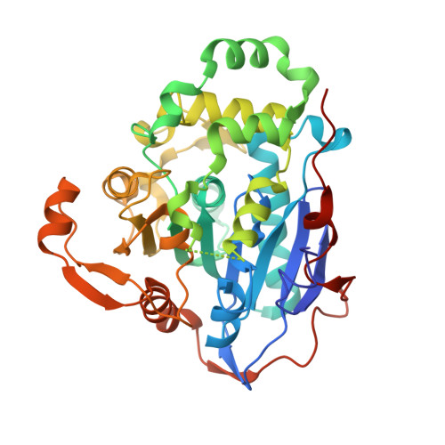
Polyhydroxyalkanoate (PHA) is a promising candidate for use as an alternative bioplastic to replace petroleum-based plastics. Our understanding of PHA synthase PhaC is poor due to the paucity of available three-dimensional structural information. Here we present a high-resolution crystal structure of the catalytic domain of PhaC from Chromobacterium sp. USM2, PhaC Cs -CAT. The structure shows that PhaC Cs -CAT forms an α/β hydrolase fold comprising α/β core and CAP subdomains. The active site containing Cys291, Asp447 and His477 is located at the bottom of the cavity, which is filled with water molecules and is covered by the partly disordered CAP subdomain. We designated our structure as the closed form, which is distinct from the recently reported catalytic domain from Cupriavidus necator (PhaC Cn -CAT). Structural comparison showed PhaC Cn -CAT adopting a partially open form maintaining a narrow substrate access channel to the active site, but no product egress. PhaC Cs -CAT forms a face-to-face dimer mediated by the CAP subdomains. This arrangement of the dimer is also distinct from that of the PhaC Cn -CAT dimer. These findings suggest that the CAP subdomain should undergo a conformational change during catalytic activity that involves rearrangement of the dimer to facilitate substrate entry and product formation and egress from the active site.
Organizational Affiliation:
Structural Biology Laboratory, Nara Institute of Science and Technology, 8916-5 Takayama, Ikoma, Nara, 630-0192, Japan.