5X5L

Crystal structure of response regulator AdeR DNA binding domain in complex with an intercistronic region


Experimental Data Snapshot

  • Method: X-RAY DIFFRACTION
  • Resolution: 2.75 Å
  • R-Value Free: 0.296 
  • R-Value Work: 0.278 
  • R-Value Observed: 0.279 

wwPDB Validation   3D Report Full Report


This is version 1.2 of the entry. See complete history


Literature

Mechanistic insight into how multidrug resistant Acinetobacter baumannii response regulator AdeR recognizes an intercistronic region.

Wen, Y.Ouyang, Z.Yu, Y.Zhou, X.Pei, Y.Devreese, B.Higgins, P.G.Zheng, F.

(2017) Nucleic Acids Res 45: 9773-9787

  • DOI: https://doi.org/10.1093/nar/gkx624
  • Primary Citation of Related Structures:  
    5X5J, 5X5L, 5XJP

  • PubMed Abstract: 

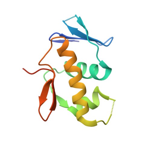
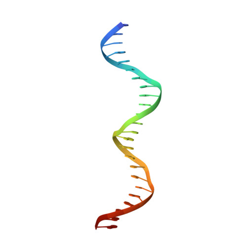
    AdeR-AdeS is a two-component regulatory system, which controls expression of the adeABC efflux pump involved in Acinetobacter baumannii multidrug resistance. AdeR is a response regulator consisting of an N-terminal receiver domain and a C-terminal DNA-binding-domain. AdeR binds to a direct-repeat DNA in the intercistronic region between adeR and adeABC. We demonstrate a markedly high affinity binding between unphosphorylated AdeR and DNA with a dissociation constant of 20 nM. In addition, we provide a 2.75 Å crystal structure of AdeR DNA-binding-domain complexed with the intercistronic DNA. This structure shows that the α3 and β hairpin formed by β5-β6 interacts with the major and minor groove of the DNA, which in turn leads to the introduction of a bend. The AdeR receiver domain structure revealed a dimerization motif mediated by a gearwheel-like structure involving the D108F109-R122 motif through cation π stack interaction. The structure of AdeR receiver domain bound with magnesium indicated a conserved Glu19Asp20-Asp63 magnesium-binding motif, and revealed that the potential phosphorylation site Asp63OD1 forms a hydrogen bond with Lys112. We thus dissected the mechanism of how AdeR recognizes the intercistronic DNA, which leads to a diverse mode of response regulation. Unlocking the AdeRS mechanism provides ways to circumvent A. baumannii antibiotic resistance.


  • Organizational Affiliation

    Center for Translational Medicine, The Key Laboratory of Biomedical Information Engineering of Ministry of Education, School of Life Science and Technology, Xi'an Jiaotong University, Xi'an 710061, China.


Macromolecules

Find similar proteins by:  (by identity cutoff)  |  3D Structure
Entity ID: 1
MoleculeChains Sequence LengthOrganismDetailsImage
AdeR109Acinetobacter baumanniiMutation(s): 0 
Gene Names: adeR
UniProt
Find proteins for E1A0Z5 (Acinetobacter baumannii)
Explore E1A0Z5 
Go to UniProtKB:  E1A0Z5
Entity Groups  
Sequence Clusters30% Identity50% Identity70% Identity90% Identity95% Identity100% Identity
UniProt GroupE1A0Z5
Sequence Annotations
Expand
  • Reference Sequence

Find similar nucleic acids by:  Sequence   |   3D Structure  

Entity ID: 2
MoleculeChains LengthOrganismImage
DNA (5'-D(P*TP*AP*AP*AP*GP*TP*GP*TP*GP*GP*AP*GP*TP*AP*AP*GP*TP*GP*TP*GP*GP*AP*GP*A)-3')G [auth C],
I [auth F]
25synthetic construct
Sequence Annotations
Expand
  • Reference Sequence

Find similar nucleic acids by:  Sequence   |   3D Structure  

Entity ID: 3
MoleculeChains LengthOrganismImage
DNA (5'-D(P*TP*CP*TP*CP*CP*AP*CP*AP*CP*TP*TP*AP*CP*TP*CP*CP*AP*CP*AP*CP*TP*TP*TP*A)-3')H [auth D],
J [auth G]
25synthetic construct
Sequence Annotations
Expand
  • Reference Sequence
Experimental Data & Validation

Experimental Data

  • Method: X-RAY DIFFRACTION
  • Resolution: 2.75 Å
  • R-Value Free: 0.296 
  • R-Value Work: 0.278 
  • R-Value Observed: 0.279 
  • Space Group: P 1
Unit Cell:
Length ( Å )Angle ( ˚ )
a = 55.51α = 101.96
b = 71.38β = 104.53
c = 78.36γ = 101.86
Software Package:
Software NamePurpose
REFMACrefinement
XDSdata reduction
XDSdata scaling
MOLREPphasing

Structure Validation

View Full Validation Report


Entry History & Funding Information

Deposition Data

  • Released Date: 2017-08-30 
  • Deposition Author(s): Wen, Y.

Funding OrganizationLocationGrant Number
National Natural Science Foundation of ChinaChinaNO.31500051

Revision History  (Full details and data files)

  • Version 1.0: 2017-08-30
    Type: Initial release
  • Version 1.1: 2017-12-13
    Changes: Database references
  • Version 1.2: 2023-11-22
    Changes: Data collection, Database references, Refinement description