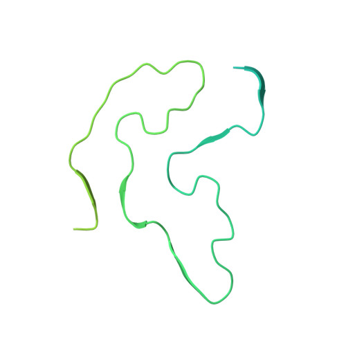
5W3N

Molecular structure of FUS low sequence complexity domain protein fibrils


Experimental Data Snapshot

  • Method: SOLID-STATE NMR
  • Conformers Calculated: 4928 
  • Conformers Submitted: 20 
  • Selection Criteria: structures with no violations, lowest energy, and derived from one of 44 independent calculations 

wwPDB Validation   3D Report Full Report


This is version 1.4 of the entry. See complete history


Literature

Structure of FUS Protein Fibrils and Its Relevance to Self-Assembly and Phase Separation of Low-Complexity Domains.

Murray, D.T.Kato, M.Lin, Y.Thurber, K.R.Hung, I.McKnight, S.L.Tycko, R.

(2017) Cell 171: 615-627.e16

  • DOI: https://doi.org/10.1016/j.cell.2017.08.048
  • Primary Citation of Related Structures:  
    5W3N

  • PubMed Abstract: 

    Polymerization and phase separation of proteins containing low-complexity (LC) domains are important factors in gene expression, mRNA processing and trafficking, and localization of translation. We have used solid-state nuclear magnetic resonance methods to characterize the molecular structure of self-assembling fibrils formed by the LC domain of the fused in sarcoma (FUS) RNA-binding protein. From the 214-residue LC domain of FUS (FUS-LC), a segment of only 57 residues forms the fibril core, while other segments remain dynamically disordered. Unlike pathogenic amyloid fibrils, FUS-LC fibrils lack hydrophobic interactions within the core and are not polymorphic at the molecular structural level. Phosphorylation of core-forming residues by DNA-dependent protein kinase blocks binding of soluble FUS-LC to FUS-LC hydrogels and dissolves phase-separated, liquid-like FUS-LC droplets. These studies offer a structural basis for understanding LC domain self-assembly, phase separation, and regulation by post-translational modification.


  • Organizational Affiliation

    Laboratory of Chemical Physics, National Institute of Diabetes and Digestive and Kidney Diseases, National Institutes of Health, Bethesda, MD 20892-0520, USA; Postdoctoral Research Associate Program, National Institute of General Medical Sciences, National Institutes of Health, Bethesda, MD 20892-6200, USA.


Macromolecules
Find similar proteins by:  (by identity cutoff)  |  3D Structure
Entity ID: 1
MoleculeChains Sequence LengthOrganismDetailsImage
RNA-binding protein FUS
A, B, C, D, E
A, B, C, D, E, F, G, H, I
241Homo sapiensMutation(s): 0 
Gene Names: FUSTLS
UniProt & NIH Common Fund Data Resources
Find proteins for P35637 (Homo sapiens)
Explore P35637 
Go to UniProtKB:  P35637
PHAROS:  P35637
GTEx:  ENSG00000089280 
Entity Groups  
Sequence Clusters30% Identity50% Identity70% Identity90% Identity95% Identity100% Identity
UniProt GroupP35637
Sequence Annotations
Expand
  • Reference Sequence
Experimental Data & Validation

Experimental Data

  • Method: SOLID-STATE NMR
  • Conformers Calculated: 4928 
  • Conformers Submitted: 20 
  • Selection Criteria: structures with no violations, lowest energy, and derived from one of 44 independent calculations 

Structure Validation

View Full Validation Report


Entry History & Funding Information

Deposition Data


Funding OrganizationLocationGrant Number
National Institutes of Health/National Institute of General Medical Sciences (NIH/NIGMS)United States5U01GM107623-03
National Institutes of Health/National Institute of General Medical Sciences (NIH/NIGMS)United States1FI2GM117604-01

Revision History  (Full details and data files)

  • Version 1.0: 2017-09-27
    Type: Initial release
  • Version 1.1: 2017-10-18
    Changes: Author supporting evidence
  • Version 1.2: 2019-04-10
    Changes: Data collection, Database references
  • Version 1.3: 2020-01-01
    Changes: Author supporting evidence, Data collection
  • Version 1.4: 2023-06-14
    Changes: Database references, Other