5VKQ

Structure of a mechanotransduction ion channel Drosophila NOMPC in nanodisc


Experimental Data Snapshot

  • Method: ELECTRON MICROSCOPY
  • Resolution: 3.55 Å
  • Aggregation State: PARTICLE 
  • Reconstruction Method: SINGLE PARTICLE 

wwPDB Validation   3D Report Full Report


This is version 1.7 of the entry. See complete history


Literature

Electron cryo-microscopy structure of the mechanotransduction channel NOMPC.

Jin, P.Bulkley, D.Guo, Y.Zhang, W.Guo, Z.Huynh, W.Wu, S.Meltzer, S.Cheng, T.Jan, L.Y.Jan, Y.N.Cheng, Y.

(2017) Nature 547: 118-122

  • DOI: https://doi.org/10.1038/nature22981
  • Primary Citation of Related Structures:  
    5VKQ

  • PubMed Abstract: 

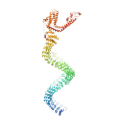
    Mechanosensory transduction for senses such as proprioception, touch, balance, acceleration, hearing and pain relies on mechanotransduction channels, which convert mechanical stimuli into electrical signals in specialized sensory cells. How force gates mechanotransduction channels is a central question in the field, for which there are two major models. One is the membrane-tension model: force applied to the membrane generates a change in membrane tension that is sufficient to gate the channel, as in the bacterial MscL channel and certain eukaryotic potassium channels. The other is the tether model: force is transmitted via a tether to gate the channel. The transient receptor potential (TRP) channel NOMPC is important for mechanosensation-related behaviours such as locomotion, touch and sound sensation across different species including Caenorhabditis elegans, Drosophila and zebrafish. NOMPC is the founding member of the TRPN subfamily, and is thought to be gated by tethering of its ankyrin repeat domain to microtubules of the cytoskeleton. Thus, a goal of studying NOMPC is to reveal the underlying mechanism of force-induced gating, which could serve as a paradigm of the tether model. NOMPC fulfils all the criteria that apply to mechanotransduction channels and has 29 ankyrin repeats, the largest number among TRP channels. A key question is how the long ankyrin repeat domain is organized as a tether that can trigger channel gating. Here we present a de novo atomic structure of Drosophila NOMPC determined by single-particle electron cryo-microscopy. Structural analysis suggests that the ankyrin repeat domain of NOMPC resembles a helical spring, suggesting its role of linking mechanical displacement of the cytoskeleton to the opening of the channel. The NOMPC architecture underscores the basis of translating mechanical force into an electrical signal within a cell.


  • Organizational Affiliation

    Department of Physiology, University of California, San Francisco, California 94158, USA.


Macromolecules
Find similar proteins by:  (by identity cutoff)  |  3D Structure
Entity ID: 1
MoleculeChains Sequence LengthOrganismDetailsImage
No mechanoreceptor potential C isoform L
A, B, C, D
1,732Drosophila melanogasterMutation(s): 0 
Gene Names: nompCCG11020Dmel_CG11020
Membrane Entity: Yes 
UniProt
Find proteins for E0A9E1 (Drosophila melanogaster)
Explore E0A9E1 
Go to UniProtKB:  E0A9E1
Entity Groups  
Sequence Clusters30% Identity50% Identity70% Identity90% Identity95% Identity100% Identity
UniProt GroupE0A9E1
Sequence Annotations
Expand
  • Reference Sequence
Small Molecules
Ligands 1 Unique
IDChains Name / Formula / InChI Key2D Diagram3D Interactions
PCF
Query on PCF

Download Ideal Coordinates CCD File 
AA [auth C]
BA [auth C]
CA [auth D]
DA [auth D]
E [auth A]
AA [auth C],
BA [auth C],
CA [auth D],
DA [auth D],
E [auth A],
EA [auth D],
F [auth A],
FA [auth D],
G [auth A],
GA [auth D],
H [auth A],
HA [auth D],
I [auth A],
IA [auth D],
J [auth A],
JA [auth D],
K [auth A],
L [auth A],
M [auth B],
N [auth B],
O [auth B],
P [auth B],
Q [auth B],
R [auth B],
S [auth B],
T [auth B],
U [auth C],
V [auth C],
W [auth C],
X [auth C],
Y [auth C],
Z [auth C]
1,2-DIACYL-SN-GLYCERO-3-PHOSHOCHOLINE
C40 H80 N O8 P
KILNVBDSWZSGLL-KXQOOQHDSA-N
Experimental Data & Validation

Experimental Data

  • Method: ELECTRON MICROSCOPY
  • Resolution: 3.55 Å
  • Aggregation State: PARTICLE 
  • Reconstruction Method: SINGLE PARTICLE 
EM Software:
TaskSoftware PackageVersion
RECONSTRUCTIONRELION1.4
MODEL REFINEMENTPHENIXdev-2608

Structure Validation

View Full Validation Report


Entry History & Funding Information

Deposition Data


Funding OrganizationLocationGrant Number
National Institutes of Health/National Institute of General Medical Sciences (NIH/NIGMS)United StatesR01GM098672
National Institutes of Health/Office of the DirectorUnited StatesS10OD020054
National Institutes of Health/National Institute of Neurological Disorders and Stroke (NIH/NINDS)United StatesR01NS069229
National Institutes of Health/National Institute of Neurological Disorders and Stroke (NIH/NINDS)United States5R37NS040929
National Institutes of Health/National Institute of Neurological Disorders and Stroke (NIH/NINDS)United States1R35NS097227
National Institutes of Health/National Institute of General Medical Sciences (NIH/NIGMS)United StatesR01GM098672
National Institutes of Health/Office of the DirectorUnited StatesS10OD020054

Revision History  (Full details and data files)

  • Version 1.0: 2017-06-28
    Type: Initial release
  • Version 1.1: 2017-07-05
    Changes: Database references
  • Version 1.2: 2017-07-12
    Changes: Database references
  • Version 1.3: 2017-07-19
    Changes: Database references
  • Version 1.4: 2017-09-20
    Changes: Author supporting evidence, Data collection, Experimental preparation
  • Version 1.5: 2019-11-06
    Changes: Data collection, Other
  • Version 1.6: 2019-12-18
    Changes: Author supporting evidence
  • Version 1.7: 2024-03-13
    Changes: Data collection, Database references