Structural insights into the interaction of the conserved mammalian proteins GAPR-1 and Beclin 1, a key autophagy protein.
Li, Y., Zhao, Y., Su, M., Glover, K., Chakravarthy, S., Colbert, C.L., Levine, B., Sinha, S.C.(2017) Acta Crystallogr D Struct Biol 73: 775-792
- PubMed: 28876241
- DOI: https://doi.org/10.1107/S2059798317011822
- Primary Citation of Related Structures:
5VHG - PubMed Abstract:
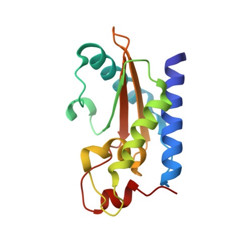
Mammalian Golgi-associated plant pathogenesis-related protein 1 (GAPR-1) is a negative autophagy regulator that binds Beclin 1, a key component of the autophagosome nucleation complex. Beclin 1 residues 267-284 are required for binding GAPR-1. Here, sequence analyses, structural modeling, mutagenesis combined with pull-down assays, X-ray crystal structure determination and small-angle X-ray scattering were used to investigate the Beclin 1-GAPR-1 interaction. Five conserved residues line an equatorial GAPR-1 surface groove that is large enough to bind a peptide. A model of a peptide comprising Beclin 1 residues 267-284 docked onto GAPR-1, built using the CABS-dock server, indicates that this peptide binds to this GAPR-1 groove. Mutation of the five conserved residues lining this groove, H54A/E86A/G102K/H103A/N138G, abrogates Beclin 1 binding. The 1.27 Å resolution X-ray crystal structure of this pentad mutant GAPR-1 was determined. Comparison with the wild-type (WT) GAPR-1 structure shows that the equatorial groove of the pentad mutant is shallower and more positively charged, and therefore may not efficiently bind Beclin 1 residues 267-284, which include many hydrophobic residues. Both WT and pentad mutant GAPR-1 crystallize as dimers, and in each case the equatorial groove of one subunit is partially occluded by the other subunit, indicating that dimeric GAPR-1 is unlikely to bind Beclin 1. SAXS analysis of WT and pentad mutant GAPR-1 indicates that in solution the WT forms monomers, while the pentad mutant is primarily dimeric. Thus, changes in the structure of the equatorial groove combined with the improved dimerization of pentad mutant GAPR-1 are likely to abrogate binding to Beclin 1.
Organizational Affiliation:
Department of Chemistry and Biochemistry, North Dakota State University, Fargo, ND 58108, USA.


