5V5R

Dehaloperoxidase A I9L mutant


Experimental Data Snapshot

  • Method: X-RAY DIFFRACTION
  • Resolution: 1.20 Å
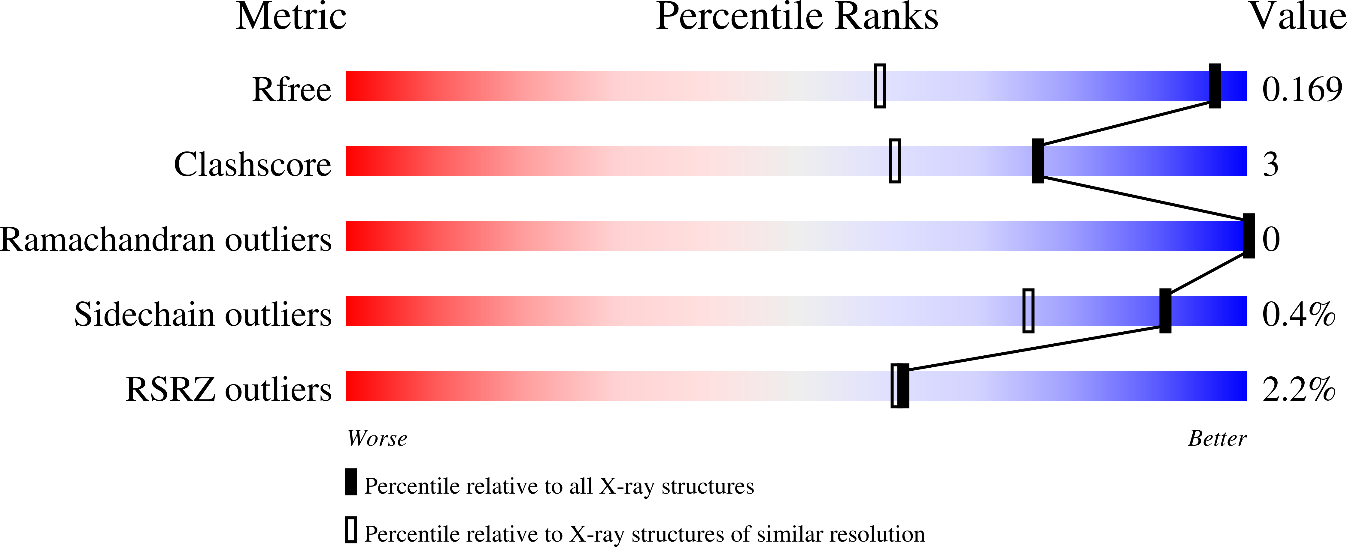
  • R-Value Free: 0.169 
  • R-Value Work: 0.139 
  • R-Value Observed: 0.140 

wwPDB Validation   3D Report Full Report


Ligand Structure Quality Assessment 


This is version 1.3 of the entry. See complete history


Literature

How nature tunes isoenzyme activity in the multifunctional catalytic globin dehaloperoxidase from Amphitrite ornata.

Carey, L.M.Gavenko, R.Svistunenko, D.A.Ghiladi, R.A.

(2018) Biochim Biophys Acta Proteins Proteom 1866: 230-241

  • DOI: https://doi.org/10.1016/j.bbapap.2017.11.004
  • Primary Citation of Related Structures:  
    5V5J, 5V5Q, 5V5R

  • PubMed Abstract: 

    The coelomic hemoglobin of Amphitrite ornata, termed dehaloperoxidase (DHP), is the first known multifunctional catalytic globin to possess biologically-relevant peroxidase and peroxygenase activities. Although the two isoenzymes of DHP, A and B, differ in sequence by only 5 amino acids out of 137 residues, DHP B consistently exhibits a greater activity than isoenzyme A. To delineate the contributions of each amino acid substitution to the activity of either isoenzyme, the substitutions of the five amino acids were systematically investigated, individually and in combination, using 22 mutants. Biochemical assays and mechanistic studies demonstrated that the mutants that only contained the I9L substitution showed increased i) k cat values (peroxidase activity), ii) 5-Br-indole conversion and binding affinity (peroxygenase activity), and iii) rate of Compound ES formation (enzyme activation). Whereas the X-ray structures of the oxyferrous forms of DHP B (L9I) (1.96Å), DHP A (I9L) (1.20Å), and WT DHP B (1.81Å) showed no significant differences, UV-visible spectroscopy (A Soret /A 380 ratio) revealed that the I9L substitution increased the 5-coordinate high-spin heme population characterized by the "open" conformation (i.e., distal histidine swung out of the pocket), which likely favors substrate binding. The positioning of the distal histidine closer to the heme cofactor in the solution state also appears to facilitate activation of DHP via the Compound ES intermediate. Taken together, the studies undertaken here shed light on the structure-function relationship in dehaloperoxidase, but also help to establish the foundation for understanding how enzymatic activity can be tuned in isoenzymes of a multifunctional catalytic globin.


  • Organizational Affiliation

    Department of Chemistry, North Carolina State University, Raleigh, NC 27695-8204, United States.


Macromolecules
Find similar proteins by:  (by identity cutoff)  |  3D Structure
Entity ID: 1
MoleculeChains Sequence LengthOrganismDetailsImage
Dehaloperoxidase AA [auth B],
B [auth A]
137Amphitrite ornataMutation(s): 1 
UniProt
Find proteins for Q9NAV8 (Amphitrite ornata)
Explore Q9NAV8 
Go to UniProtKB:  Q9NAV8
Entity Groups  
Sequence Clusters30% Identity50% Identity70% Identity90% Identity95% Identity100% Identity
UniProt GroupQ9NAV8
Sequence Annotations
Expand
  • Reference Sequence
Small Molecules
Ligands 4 Unique
IDChains Name / Formula / InChI Key2D Diagram3D Interactions
HEM
Query on HEM

Download Ideal Coordinates CCD File 
C [auth B],
F [auth A]
PROTOPORPHYRIN IX CONTAINING FE
C34 H32 Fe N4 O4
KABFMIBPWCXCRK-RGGAHWMASA-L
SO4
Query on SO4

Download Ideal Coordinates CCD File 
G [auth A],
H [auth A]
SULFATE ION
O4 S
QAOWNCQODCNURD-UHFFFAOYSA-L
GOL
Query on GOL

Download Ideal Coordinates CCD File 
E [auth B]GLYCEROL
C3 H8 O3
PEDCQBHIVMGVHV-UHFFFAOYSA-N
OXY
Query on OXY

Download Ideal Coordinates CCD File 
D [auth B],
I [auth A]
OXYGEN MOLECULE
O2
MYMOFIZGZYHOMD-UHFFFAOYSA-N
Experimental Data & Validation

Experimental Data

  • Method: X-RAY DIFFRACTION
  • Resolution: 1.20 Å
  • R-Value Free: 0.169 
  • R-Value Work: 0.139 
  • R-Value Observed: 0.140 
  • Space Group: P 21 21 21
Unit Cell:
Length ( Å )Angle ( ˚ )
a = 57.665α = 90
b = 67.423β = 90
c = 68.91γ = 90
Software Package:
Software NamePurpose
PHENIXrefinement
HKL-2000data reduction
HKL-2000data scaling
PHASERphasing

Structure Validation

View Full Validation Report


Ligand Structure Quality Assessment 


Entry History & Funding Information

Deposition Data


Funding OrganizationLocationGrant Number
National Science Foundation (NSF, United States)United StatesCHE-1150709

Revision History  (Full details and data files)

  • Version 1.0: 2018-03-21
    Type: Initial release
  • Version 1.1: 2019-04-03
    Changes: Data collection, Database references
  • Version 1.2: 2019-11-27
    Changes: Author supporting evidence
  • Version 1.3: 2023-10-04
    Changes: Data collection, Database references, Derived calculations, Refinement description