5TRB

Crystal structure of the RNF20 RING domain


Experimental Data Snapshot

  • Method: X-RAY DIFFRACTION
  • Resolution: 1.80 Å
  • R-Value Free: 0.235 
  • R-Value Work: 0.216 
  • R-Value Observed: 0.217 

wwPDB Validation   3D Report Full Report


This is version 1.1 of the entry. See complete history


Literature

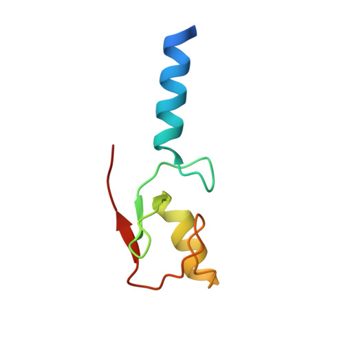
Structure and Function of the RING Domains of RNF20 and RNF40, Dimeric E3 Ligases that Monoubiquitylate Histone H2B.

Foglizzo, M.Middleton, A.J.Day, C.L.

(2016) J Mol Biol 428: 4073-4086

  • DOI: https://doi.org/10.1016/j.jmb.2016.07.025
  • Primary Citation of Related Structures:  
    5TRB

  • PubMed Abstract: 

    Monoubiquitylation of histone H2B is a post-translational mark that plays key roles in regulation of transcription and genome stability. In humans, attachment of ubiquitin to lysine 120 of histone H2B depends on the activity of the E2 ubiquitin-conjugating enzyme, Ube2B, and the really interesting new gene (RING) E3 ligases, RING finger protein (RNF) 20 and RNF40. To better understand the molecular basis of this modification, we have solved the crystal structure of the RNF20 RING domain and show that it is a homodimer that specifically interacts with the Ube2B~Ub conjugate. By mutating residues at the E3-E2 and E3-ubiquitin interfaces, we identify key contacts required for interaction of the RNF20 RING domain with the Ube2B~Ub conjugate. These mutants were used to generate a structure-based model of the RNF20-Ube2B~Ub complex that reveals differences from other RING-E2~Ub complexes, and suggests how the RNF20-Ube2B~Ub complex might interact with its nucleosomal substrate. Additionally, we show that the RING domains of RNF20 and RNF40 can form a stable heterodimer that is active. Together, our studies provide new insights into the mechanisms that regulate RNF20-mediated ubiquitin transfer from Ube2B.


  • Organizational Affiliation

    Biochemistry Department, Otago School of Medical Sciences, University of Otago, Dunedin 9054, New Zealand.


Macromolecules
Find similar proteins by:  (by identity cutoff)  |  3D Structure
Entity ID: 1
MoleculeChains Sequence LengthOrganismDetailsImage
E3 ubiquitin-protein ligase BRE1A75Homo sapiensMutation(s): 0 
Gene Names: RNF20BRE1A
EC: 6.3.2
UniProt & NIH Common Fund Data Resources
Find proteins for Q5VTR2 (Homo sapiens)
Explore Q5VTR2 
Go to UniProtKB:  Q5VTR2
PHAROS:  Q5VTR2
GTEx:  ENSG00000155827 
Entity Groups  
Sequence Clusters30% Identity50% Identity70% Identity90% Identity95% Identity100% Identity
UniProt GroupQ5VTR2
Sequence Annotations
Expand
  • Reference Sequence
Experimental Data & Validation

Experimental Data

  • Method: X-RAY DIFFRACTION
  • Resolution: 1.80 Å
  • R-Value Free: 0.235 
  • R-Value Work: 0.216 
  • R-Value Observed: 0.217 
  • Space Group: I 21 21 21
Unit Cell:
Length ( Å )Angle ( ˚ )
a = 34.83α = 90
b = 61.29β = 90
c = 78.67γ = 90
Software Package:
Software NamePurpose
SCALAdata scaling
PHENIXrefinement
PDB_EXTRACTdata extraction
PHENIXphasing
MOSFLMdata reduction

Structure Validation

View Full Validation Report


Entry History & Funding Information

Deposition Data


Funding OrganizationLocationGrant Number
Marsden FundNew Zealand--

Revision History  (Full details and data files)

  • Version 1.0: 2016-11-02
    Type: Initial release
  • Version 1.1: 2024-03-06
    Changes: Data collection, Database references, Derived calculations