Refined 1.83 A structure of trypanosomal triosephosphate isomerase crystallized in the presence of 2.4 M-ammonium sulphate. A comparison with the structure of the trypanosomal triosephosphate isomerase-glycerol-3-phosphate complex.
Wierenga, R.K., Noble, M.E., Vriend, G., Nauche, S., Hol, W.G.(1991) J Mol Biol 220: 995-1015
- PubMed: 1880808
- DOI: https://doi.org/10.1016/0022-2836(91)90368-g
- Primary Citation of Related Structures:
5TIM - PubMed Abstract:
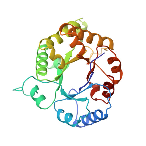
Triosephosphate isomerase (TIM) is a dimeric glycolytic enzyme. TIM from Trypanosoma brucei brucei has been crystallized at pH 7.0 in 2.4 M-ammonium sulphate. The well-diffracting crystals have one dimer per asymmetric unit. The structure has been refined at 1.83 A resolution with an R-factor of 18.3% for all data between 6 A and 1.83 A (37,568 reflections). The model consists of 3778 protein atoms and 297 solvent atoms. Subunit 1 is involved in considerably more crystal contacts than subunit 2. Correlated with these differences in crystal packing is the observation that only in the active site of subunit 2 is a sulphate ion bound. Furthermore, significant differences with respect to structure and flexibility are observed in three loops near the active site. In particular, there is a 7 A positional difference of the tip of the flexible loop (loop 6) when comparing subunit 1 and subunit 2. Also, the neighbouring loops (loop 5 and loop 7) have significantly different conformations and flexibility. In subunit 1, loop 6 is in an "open" conformation, in subunit 2, loop 6 is in an "almost closed" conformation. Only in the presence of a phosphate-containing ligand, such as glycerol-3-phosphate, does loop 6 take up the "closed" conformation. Loop 6 and loop 7 (and also to some extent loop 5) are rather flexible in the almost closed conformation, but well defined in the open and closed conformations. The closing of loop 6 (167 to 180), as observed in the almost closed conformation, slightly changes the main-chain conformation of the catalytic glutamate, Glu167, leading to a change of the chi 1 angle of this residue from approximately -60 degrees to approximately 60 degrees and the weakening of the hydrogen bonds between its polar side-chain atoms and Ser96. In the closed conformation, in the presence of glycerol-3-phosphate, the main-chain atoms of Glu167 remain in the same position as in the almost closed conformation, but the side-chain has rotated around the CA-CB bond changing chi 1 from approximately 60 degrees to approximately -60 degrees. In this new position the hydrogen bonding to Ser96 is completely lost and also a water-mediated salt bridge between OE2(Glu167) and NE(Arg99) is lost. Comparison of the two independently refined subunits, showed that the root-mean-square deviation for all 249 CA atoms is 0.9 A; for the CA atoms of the beta-strands this is only 0.2 A. The average B-factor for all subunit 1 and subunit 2 atoms is 20 A2 and 25 A2, respectively.(ABSTRACT TRUNCATED AT 400 WORDS)
Organizational Affiliation:
European Molecular Biology Laboratory, Heidelberg, Germany.