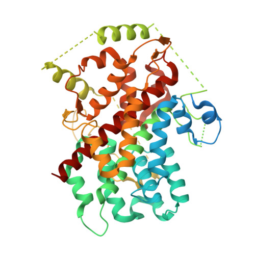
The apo-structure of the leucine sensor Sestrin2 is still elusive.
Saxton, R.A., Knockenhauer, K.E., Schwartz, T.U., Sabatini, D.M.(2016) Sci Signal 9: ra92-ra92
- PubMed: 27649739
- DOI: https://doi.org/10.1126/scisignal.aah4497
- Primary Citation of Related Structures:
5T0N - PubMed Abstract:
Sestrin2 is a GATOR2-interacting protein that directly binds leucine and is required for the inhibition of mTORC1 under leucine deprivation, indicating that it is a leucine sensor for the mTORC1 pathway. We recently reported the structure of Sestrin2 in complex with leucine [Protein Data Bank (PDB) ID, 5DJ4] and demonstrated that mutations in the leucine-binding pocket that alter the affinity of Sestrin2 for leucine result in a corresponding change in the leucine sensitivity of mTORC1 in cells. A lower resolution structure of human Sestrin2 (PDB ID, 5CUF), which was crystallized in the absence of exogenous leucine, showed Sestrin2 to be in a nearly identical conformation as the leucine-bound structure. On the basis of this observation, it has been argued that leucine binding does not affect the conformation of Sestrin2 and that Sestrin2 may not be a sensor for leucine. We show that simple analysis of the reported "apo"-Sestrin2 structure reveals the clear presence of prominent, unmodeled electron density in the leucine-binding pocket that exactly accommodates the leucine observed in the higher resolution structure. Refining the reported apo-structure with leucine eliminated the large Fobs-Fcalc difference density at this position and improved the working and free R factors of the model. Consistent with this result, our own structure of Sestrin2 crystallized in the absence of exogenous leucine also contained electron density that is best explained by leucine. Thus, the structure of apo-Sestrin2 remains elusive.
Organizational Affiliation:
Department of Biology, Massachusetts Institute of Technology, Cambridge, MA 02139, USA. Whitehead Institute for Biomedical Research, 9 Cambridge Center, Cambridge, MA 02142, USA. Howard Hughes Medical Institute, Cambridge, MA 02139, USA. Koch Institute for Integrative Cancer Research, 77 Massachusetts Avenue, Cambridge, MA 02139, USA. Broad Institute of Harvard and Massachusetts Institute of Technology, 415 Main Street, Cambridge, MA 02142, USA.