5SV7

The Crystal structure of a chaperone


Experimental Data Snapshot

  • Method: X-RAY DIFFRACTION
  • Resolution: 3.21 Å
  • R-Value Free: 0.322 
  • R-Value Work: 0.271 
  • R-Value Observed: 0.273 

wwPDB Validation   3D Report Full Report


This is version 1.3 of the entry. See complete history


Literature

The ER stress sensor PERK luminal domain functions as a molecular chaperone to interact with misfolded proteins.

Wang, P.Li, J.Sha, B.

(2016) Acta Crystallogr D Struct Biol 72: 1290-1297

  • DOI: https://doi.org/10.1107/S2059798316018064
  • Primary Citation of Related Structures:  
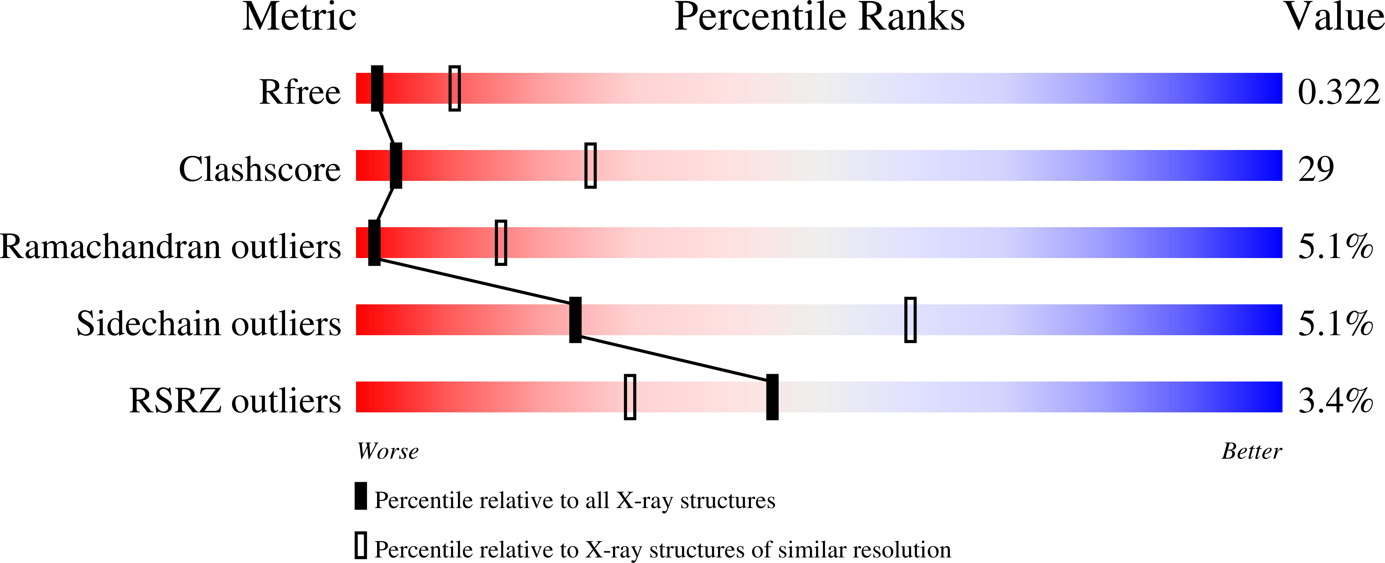
    5SV7

  • PubMed Abstract: 

    PERK is one of the major sensor proteins which can detect the protein-folding imbalance generated by endoplasmic reticulum (ER) stress. It remains unclear how the sensor protein PERK is activated by ER stress. It has been demonstrated that the PERK luminal domain can recognize and selectively interact with misfolded proteins but not native proteins. Moreover, the PERK luminal domain may function as a molecular chaperone to directly bind to and suppress the aggregation of a number of misfolded model proteins. The data strongly support the hypothesis that the PERK luminal domain can interact directly with misfolded proteins to induce ER stress signaling. To illustrate the mechanism by which the PERK luminal domain interacts with misfolded proteins, the crystal structure of the human PERK luminal domain was determined to 3.2 Å resolution. Two dimers of the PERK luminal domain constitute a tetramer in the asymmetric unit. Superimposition of the PERK luminal domain molecules indicated that the β-sandwich domain could adopt multiple conformations. It is hypothesized that the PERK luminal domain may utilize its flexible β-sandwich domain to recognize and interact with a broad range of misfolded proteins.


  • Organizational Affiliation

    Department of Cell, Developmental and Integrative Biology (CDIB), University of Alabama at Birmingham, Birmingham, AL 35294, USA.


Macromolecules
Find similar proteins by:  (by identity cutoff)  |  3D Structure
Entity ID: 1
MoleculeChains Sequence LengthOrganismDetailsImage
Eukaryotic translation initiation factor 2-alpha kinase 3
A, B, C, D
326Homo sapiensMutation(s): 0 
Gene Names: EIF2AK3PEKPERK
EC: 2.7.11.1
UniProt & NIH Common Fund Data Resources
Find proteins for Q9NZJ5 (Homo sapiens)
Explore Q9NZJ5 
Go to UniProtKB:  Q9NZJ5
PHAROS:  Q9NZJ5
GTEx:  ENSG00000172071 
Entity Groups  
Sequence Clusters30% Identity50% Identity70% Identity90% Identity95% Identity100% Identity
UniProt GroupQ9NZJ5
Sequence Annotations
Expand
  • Reference Sequence
Experimental Data & Validation

Experimental Data

  • Method: X-RAY DIFFRACTION
  • Resolution: 3.21 Å
  • R-Value Free: 0.322 
  • R-Value Work: 0.271 
  • R-Value Observed: 0.273 
  • Space Group: P 32
Unit Cell:
Length ( Å )Angle ( ˚ )
a = 163.913α = 90
b = 163.913β = 90
c = 63.076γ = 120
Software Package:
Software NamePurpose
PHENIXrefinement
HKL-2000data reduction
HKL-2000data scaling
PHASERphasing

Structure Validation

View Full Validation Report


Entry History & Funding Information

Deposition Data


Funding OrganizationLocationGrant Number
National Institutes of Health/National Institute of General Medical Sciences (NIH/NIGMS)United StatesR01 GM080261

Revision History  (Full details and data files)

  • Version 1.0: 2017-03-01
    Type: Initial release
  • Version 1.1: 2017-09-13
    Changes: Author supporting evidence
  • Version 1.2: 2022-04-13
    Changes: Author supporting evidence, Database references
  • Version 1.3: 2023-10-04
    Changes: Data collection, Refinement description