5SUK

G6P bound activated state of yeast glycogen synthase 2


Experimental Data Snapshot

  • Method: X-RAY DIFFRACTION
  • Resolution: 2.88 Å
  • R-Value Free: 0.260 
  • R-Value Work: 0.219 
  • R-Value Observed: 0.221 

wwPDB Validation   3D Report Full Report


Ligand Structure Quality Assessment 


This is version 1.3 of the entry. See complete history


Literature

Redox Switch for the Inhibited State of Yeast Glycogen Synthase Mimics Regulation by Phosphorylation.

Mahalingan, K.K.Baskaran, S.DePaoli-Roach, A.A.Roach, P.J.Hurley, T.D.

(2017) Biochemistry 56: 179-188

  • DOI: https://doi.org/10.1021/acs.biochem.6b00884
  • Primary Citation of Related Structures:  
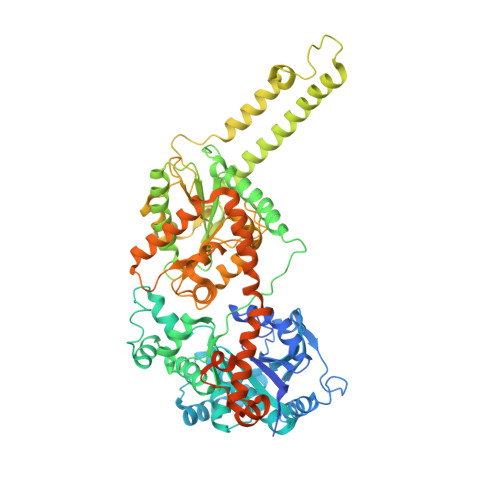
    5SUK, 5SUL

  • PubMed Abstract: 

    Glycogen synthase (GS) is the rate limiting enzyme in the synthesis of glycogen. Eukaryotic GS is negatively regulated by covalent phosphorylation and allosterically activated by glucose-6-phosphate (G-6-P). To gain structural insights into the inhibited state of the enzyme, we solved the crystal structure of yGsy2-R589A/R592A to a resolution of 3.3 Å. The double mutant has an activity ratio similar to the phosphorylated enzyme and also retains the ability to be activated by G-6-P. When compared to the 2.88 Å structure of the wild-type G-6-P activated enzyme, the crystal structure of the low-activity mutant showed that the N-terminal domain of the inhibited state is tightly held against the dimer-related interface thereby hindering acceptor access to the catalytic cleft. On the basis of these two structural observations, we developed a reversible redox regulatory feature in yeast GS by substituting cysteine residues for two highly conserved arginine residues. When oxidized, the cysteine mutant enzyme exhibits activity levels similar to the phosphorylated enzyme but cannot be activated by G-6-P. Upon reduction, the cysteine mutant enzyme regains normal activity levels and regulatory response to G-6-P activation.


  • Organizational Affiliation

    Department of Biochemistry and Molecular Biology, Indiana University School of Medicine , Indianapolis, Indiana 46202, United States.


Macromolecules
Find similar proteins by:  (by identity cutoff)  |  3D Structure
Entity ID: 1
MoleculeChains Sequence LengthOrganismDetailsImage
Glycogen [starch] synthase isoform 2
A, B, C, D
725Saccharomyces cerevisiae S288CMutation(s): 0 
Gene Names: GSY2YLR258WL8479.8
EC: 2.4.1.11
UniProt
Find proteins for P27472 (Saccharomyces cerevisiae (strain ATCC 204508 / S288c))
Explore P27472 
Go to UniProtKB:  P27472
Entity Groups  
Sequence Clusters30% Identity50% Identity70% Identity90% Identity95% Identity100% Identity
UniProt GroupP27472
Sequence Annotations
Expand
  • Reference Sequence
Experimental Data & Validation

Experimental Data

  • Method: X-RAY DIFFRACTION
  • Resolution: 2.88 Å
  • R-Value Free: 0.260 
  • R-Value Work: 0.219 
  • R-Value Observed: 0.221 
  • Space Group: I 2 2 2
Unit Cell:
Length ( Å )Angle ( ˚ )
a = 191.984α = 90
b = 204.562β = 90
c = 205.819γ = 90
Software Package:
Software NamePurpose
REFMACrefinement
HKL-2000data reduction
HKL-2000data scaling
MOLREPphasing

Structure Validation

View Full Validation Report


Ligand Structure Quality Assessment 


Entry History 

Deposition Data

Revision History  (Full details and data files)

  • Version 1.0: 2017-06-14
    Type: Initial release
  • Version 1.1: 2018-12-26
    Changes: Data collection, Database references
  • Version 1.2: 2020-07-29
    Type: Remediation
    Reason: Carbohydrate remediation
    Changes: Data collection, Derived calculations, Structure summary
  • Version 1.3: 2023-10-04
    Changes: Data collection, Database references, Refinement description, Structure summary