5OL2

The electron transferring flavoprotein/butyryl-CoA dehydrogenase complex from Clostridium difficile


Experimental Data Snapshot

  • Method: X-RAY DIFFRACTION
  • Resolution: 3.10 Å
  • R-Value Free: 0.260 
  • R-Value Work: 0.209 
  • R-Value Observed: 0.211 

wwPDB Validation   3D Report Full Report


Ligand Structure Quality Assessment 


This is version 1.1 of the entry. See complete history


Literature

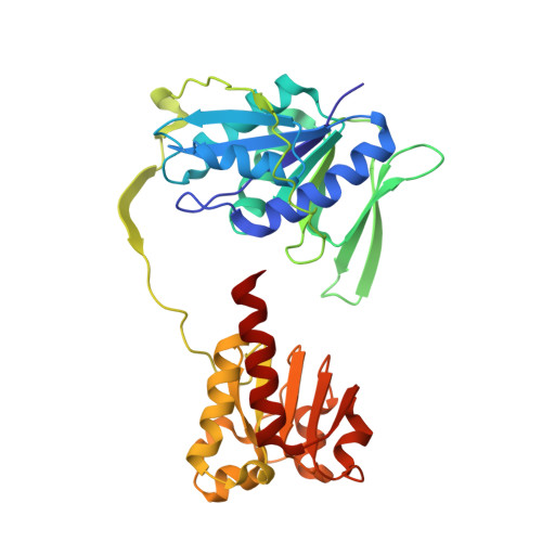
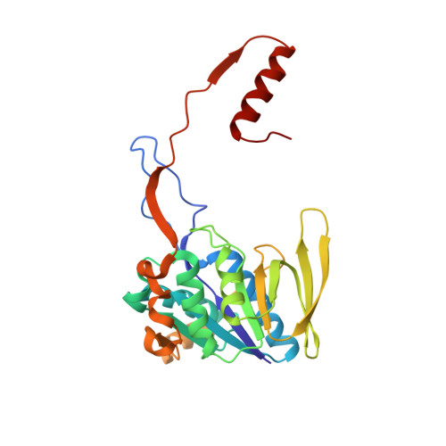
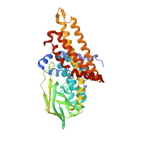
The semiquinone swing in the bifurcating electron transferring flavoprotein/butyryl-CoA dehydrogenase complex from Clostridium difficile.

Demmer, J.K.Pal Chowdhury, N.Selmer, T.Ermler, U.Buckel, W.

(2017) Nat Commun 8: 1577-1577

  • DOI: https://doi.org/10.1038/s41467-017-01746-3
  • Primary Citation of Related Structures:  
    5OL2

  • PubMed Abstract: 

    The electron transferring flavoprotein/butyryl-CoA dehydrogenase (EtfAB/Bcd) catalyzes the reduction of one crotonyl-CoA and two ferredoxins by two NADH within a flavin-based electron-bifurcating process. Here we report on the X-ray structure of the Clostridium difficile (EtfAB/Bcd) 4 complex in the dehydrogenase-conducting D-state, α-FAD (bound to domain II of EtfA) and δ-FAD (bound to Bcd) being 8 Å apart. Superimposing Acidaminococcus fermentans EtfAB onto C. difficile EtfAB/Bcd reveals a rotation of domain II of nearly 80°. Further rotation by 10° brings EtfAB into the bifurcating B-state, α-FAD and β-FAD (bound to EtfB) being 14 Å apart. This dual binding mode of domain II, substantiated by mutational studies, resembles findings in non-bifurcating EtfAB/acyl-CoA dehydrogenase complexes. In our proposed mechanism, NADH reduces β-FAD, which bifurcates. One electron goes to ferredoxin and one to α-FAD, which swings over to reduce δ-FAD to the semiquinone. Repetition affords a second reduced ferredoxin and δ-FADH - , which reduces crotonyl-CoA.


  • Organizational Affiliation

    Max-Planck-Institut für Biophysik, Max-von-Laue-Str. 3, 60438, Frankfurt am Main, Germany.


Macromolecules
Find similar proteins by:  (by identity cutoff)  |  3D Structure
Entity ID: 1
MoleculeChains Sequence LengthOrganismDetailsImage
Electron transfer flavoprotein large subunit
A, D
331Clostridioides difficileMutation(s): 0 
Gene Names: etfAetfA_4BN1095_140023BN1096_550022BN1097_530021SAMEA3374989_03962SAMEA3375004_04103SAMEA3375059_03747
UniProt
Find proteins for A0A125V455 (Clostridioides difficile ATCC 9689 = DSM 1296)
Explore A0A125V455 
Go to UniProtKB:  A0A125V455
Entity Groups  
Sequence Clusters30% Identity50% Identity70% Identity90% Identity95% Identity100% Identity
UniProt GroupA0A125V455
Sequence Annotations
Expand
  • Reference Sequence
Find similar proteins by:  (by identity cutoff)  |  3D Structure
Entity ID: 2
MoleculeChains Sequence LengthOrganismDetailsImage
Electron transfer flavoprotein small subunit
B, E
260Clostridioides difficileMutation(s): 0 
Gene Names: 
UniProt
Find proteins for A0A125V3V6 (Clostridioides difficile ATCC 9689 = DSM 1296)
Explore A0A125V3V6 
Go to UniProtKB:  A0A125V3V6
Entity Groups  
Sequence Clusters30% Identity50% Identity70% Identity90% Identity95% Identity100% Identity
UniProt GroupA0A125V3V6
Sequence Annotations
Expand
  • Reference Sequence
Find similar proteins by:  (by identity cutoff)  |  3D Structure
Entity ID: 3
MoleculeChains Sequence LengthOrganismDetailsImage
Acyl-CoA dehydrogenase
C, F
378Clostridioides difficileMutation(s): 0 
Gene Names: acdAbcdBN1095_140021BN1096_550020BN1097_530019IM33_05890SAMEA3374973_01502SAMEA3374989_03964SAMEA3375004_04105SAMEA3375059_03749
EC: 1.3.8.1 (PDB Primary Data), 1.3.99 (PDB Primary Data)
UniProt
Find proteins for A0A125V4E7 (Clostridioides difficile ATCC 9689 = DSM 1296)
Explore A0A125V4E7 
Go to UniProtKB:  A0A125V4E7
Entity Groups  
Sequence Clusters30% Identity50% Identity70% Identity90% Identity95% Identity100% Identity
UniProt GroupA0A125V4E7
Sequence Annotations
Expand
  • Reference Sequence
Small Molecules
Ligands 3 Unique
IDChains Name / Formula / InChI Key2D Diagram3D Interactions
COS
Query on COS

Download Ideal Coordinates CCD File 
K [auth C],
R [auth F]
COENZYME A PERSULFIDE
C21 H36 N7 O16 P3 S2
REVPHPVBPSIEKM-IBOSZNHHSA-N
FAD
Query on FAD

Download Ideal Coordinates CCD File 
G [auth A]
I [auth B]
J [auth C]
M [auth D]
O [auth E]
G [auth A],
I [auth B],
J [auth C],
M [auth D],
O [auth E],
Q [auth F]
FLAVIN-ADENINE DINUCLEOTIDE
C27 H33 N9 O15 P2
VWWQXMAJTJZDQX-UYBVJOGSSA-N
CA
Query on CA

Download Ideal Coordinates CCD File 
H [auth A],
L [auth C],
N [auth D],
P [auth E],
S [auth F]
CALCIUM ION
Ca
BHPQYMZQTOCNFJ-UHFFFAOYSA-N
Experimental Data & Validation

Experimental Data

  • Method: X-RAY DIFFRACTION
  • Resolution: 3.10 Å
  • R-Value Free: 0.260 
  • R-Value Work: 0.209 
  • R-Value Observed: 0.211 
  • Space Group: I 41 2 2
Unit Cell:
Length ( Å )Angle ( ˚ )
a = 177.06α = 90
b = 177.06β = 90
c = 493.15γ = 90
Software Package:
Software NamePurpose
PHENIXrefinement
XSCALEdata scaling
PHASERphasing
PDB_EXTRACTdata extraction
XDSdata reduction

Structure Validation

View Full Validation Report


Ligand Structure Quality Assessment 


Entry History & Funding Information

Deposition Data


Funding OrganizationLocationGrant Number
Max Planck SocietyGermany--
German Research FoundationGermany--
SYNMIKROGermany--

Revision History  (Full details and data files)

  • Version 1.0: 2017-11-29
    Type: Initial release
  • Version 1.1: 2024-01-17
    Changes: Data collection, Database references, Derived calculations, Refinement description