5NV8

Structural basis for EarP-mediated arginine glycosylation of translation elongation factor EF-P


Experimental Data Snapshot

  • Method: X-RAY DIFFRACTION
  • Resolution: 2.29 Å
  • R-Value Free: 0.339 
  • R-Value Work: 0.289 
  • R-Value Observed: 0.292 

wwPDB Validation   3D Report Full Report


Ligand Structure Quality Assessment 


This is version 1.2 of the entry. See complete history


Literature

Structural Basis for EarP-Mediated Arginine Glycosylation of Translation Elongation Factor EF-P.

Krafczyk, R.Macosek, J.Jagtap, P.K.A.Gast, D.Wunder, S.Mitra, P.Jha, A.K.Rohr, J.Hoffmann-Roder, A.Jung, K.Hennig, J.Lassak, J.

(2017) mBio 8

  • DOI: https://doi.org/10.1128/mBio.01412-17
  • Primary Citation of Related Structures:  
    5NV8

  • PubMed Abstract: 

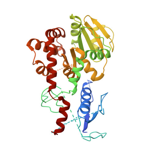
    Glycosylation is a universal strategy to posttranslationally modify proteins. The recently discovered arginine rhamnosylation activates the polyproline-specific bacterial translation elongation factor EF-P. EF-P is rhamnosylated on arginine 32 by the glycosyltransferase EarP. However, the enzymatic mechanism remains elusive. In the present study, we solved the crystal structure of EarP from Pseudomonas putida The enzyme is composed of two opposing domains with Rossmann folds, thus constituting a B pattern-type glycosyltransferase (GT-B). While dTDP-β-l-rhamnose is located within a highly conserved pocket of the C-domain, EarP recognizes the KOW-like N-domain of EF-P. Based on our data, we propose a structural model for arginine glycosylation by EarP. As EarP is essential for pathogenicity in P. aeruginosa , our study provides the basis for targeted inhibitor design. IMPORTANCE The structural and biochemical characterization of the EF-P-specific rhamnosyltransferase EarP not only provides the first molecular insights into arginine glycosylation but also lays the basis for targeted-inhibitor design against Pseudomonas aeruginosa infection.


  • Organizational Affiliation

    Center for integrated Protein Science Munich (CiPSM), Department of Biology I, Microbiology, Ludwig-Maximilians-Universität München, Munich, Germany.


Macromolecules
Find similar proteins by:  (by identity cutoff)  |  3D Structure
Entity ID: 1
MoleculeChains Sequence LengthOrganismDetailsImage
EF-P arginine 32 rhamnosyl-transferase390Pseudomonas putida KT2440Mutation(s): 0 
Gene Names: earPPP_1857
UniProt
Find proteins for Q88LS1 (Pseudomonas putida (strain ATCC 47054 / DSM 6125 / CFBP 8728 / NCIMB 11950 / KT2440))
Explore Q88LS1 
Go to UniProtKB:  Q88LS1
Entity Groups  
Sequence Clusters30% Identity50% Identity70% Identity90% Identity95% Identity100% Identity
UniProt GroupQ88LS1
Sequence Annotations
Expand
  • Reference Sequence
Small Molecules
Ligands 1 Unique
IDChains Name / Formula / InChI Key2D Diagram3D Interactions
TRH
Query on TRH

Download Ideal Coordinates CCD File 
B [auth A]2'-DEOXY-THYMIDINE-BETA-L-RHAMNOSE
C16 H26 N2 O15 P2
ZOSQFDVXNQFKBY-CGAXJHMRSA-N
Experimental Data & Validation

Experimental Data

  • Method: X-RAY DIFFRACTION
  • Resolution: 2.29 Å
  • R-Value Free: 0.339 
  • R-Value Work: 0.289 
  • R-Value Observed: 0.292 
  • Space Group: I 4
Unit Cell:
Length ( Å )Angle ( ˚ )
a = 131.68α = 90
b = 131.68β = 90
c = 46.24γ = 90
Software Package:
Software NamePurpose
PHENIXrefinement
XSCALEdata scaling
PDB_EXTRACTdata extraction
XDSdata reduction
CRANK2phasing

Structure Validation

View Full Validation Report


Ligand Structure Quality Assessment 


Entry History 

Deposition Data

Revision History  (Full details and data files)

  • Version 1.0: 2017-10-04
    Type: Initial release
  • Version 1.1: 2017-10-11
    Changes: Database references
  • Version 1.2: 2017-10-18
    Changes: Structure summary