5NIY

Signal recognition particle-docking protein FtsY


Experimental Data Snapshot

  • Method: X-RAY DIFFRACTION
  • Resolution: 1.70 Å
  • R-Value Free: 0.195 
  • R-Value Work: 0.167 
  • R-Value Observed: 0.168 

wwPDB Validation   3D Report Full Report


Ligand Structure Quality Assessment 


This is version 1.2 of the entry. See complete history


Literature

The Escherichia coli SRP Receptor Forms a Homodimer at the Membrane.

Kempf, G.Stjepanovic, G.Sloan, J.Hendricks, A.Lapouge, K.Sinning, I.

(2018) Structure 26: 1440-1450.e5

  • DOI: https://doi.org/10.1016/j.str.2018.07.008
  • Primary Citation of Related Structures:  
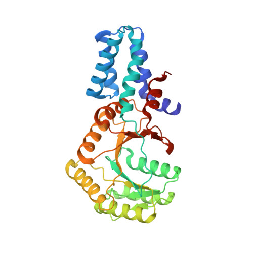
    5NIY

  • PubMed Abstract: 

    The Escherichia coli signal recognition particle (SRP) receptor, FtsY, plays a fundamental role in co-translational targeting of membrane proteins via the SRP pathway. Efficient targeting relies on membrane interaction of FtsY and heterodimerization with the SRP protein Ffh, which is driven by detachment of α helix (αN1) in FtsY. Here we show that apart from the heterodimer, FtsY forms a nucleotide-dependent homodimer on the membrane, and upon αN1 removal also in solution. Homodimerization triggers reciprocal stimulation of GTP hydrolysis and occurs in vivo. Biochemical characterization together with integrative modeling suggests that the homodimer employs the same interface as the heterodimer. Structure determination of FtsY NG+1 with GMPPNP shows that a dimerization-induced conformational switch of the γ-phosphate is conserved in Escherichia coli, filling an important gap in SRP GTPase activation. Our findings add to the current understanding of SRP GTPases and may challenge previous studies that did not consider homodimerization of FtsY.


  • Organizational Affiliation

    Heidelberg University Biochemistry Centre, Heidelberg 69120, Germany.


Macromolecules
Find similar proteins by:  (by identity cutoff)  |  3D Structure
Entity ID: 1
MoleculeChains Sequence LengthOrganismDetailsImage
Signal recognition particle-docking protein FtsY309Escherichia coliMutation(s): 0 
Gene Names: BK350_09725BK355_12030BK400_00245
UniProt
Find proteins for P10121 (Escherichia coli (strain K12))
Explore P10121 
Go to UniProtKB:  P10121
Entity Groups  
Sequence Clusters30% Identity50% Identity70% Identity90% Identity95% Identity100% Identity
UniProt GroupP10121
Sequence Annotations
Expand
  • Reference Sequence
Small Molecules
Ligands 1 Unique
IDChains Name / Formula / InChI Key2D Diagram3D Interactions
GNP
Query on GNP

Download Ideal Coordinates CCD File 
B [auth A]PHOSPHOAMINOPHOSPHONIC ACID-GUANYLATE ESTER
C10 H17 N6 O13 P3
UQABYHGXWYXDTK-UUOKFMHZSA-N
Experimental Data & Validation

Experimental Data

  • Method: X-RAY DIFFRACTION
  • Resolution: 1.70 Å
  • R-Value Free: 0.195 
  • R-Value Work: 0.167 
  • R-Value Observed: 0.168 
  • Space Group: P 1 21 1
Unit Cell:
Length ( Å )Angle ( ˚ )
a = 32.73α = 90
b = 80.64β = 95.23
c = 59.307γ = 90
Software Package:
Software NamePurpose
PHENIXrefinement
XDSdata reduction
Aimlessdata scaling
PHASERphasing

Structure Validation

View Full Validation Report


Ligand Structure Quality Assessment 


Entry History & Funding Information

Deposition Data


Funding OrganizationLocationGrant Number
German Research FoundationGermanySFB TRR83
German Research FoundationGermanyLeibniz Programm
German Research FoundationGermanyBZH/Cluster of Excellence:CellNetworks

Revision History  (Full details and data files)

  • Version 1.0: 2018-10-10
    Type: Initial release
  • Version 1.1: 2018-11-14
    Changes: Data collection, Database references, Structure summary
  • Version 1.2: 2024-01-17
    Changes: Data collection, Database references, Refinement description