5MGZ

Streptomyces Spheroides NovO (8-demethylnovbiocic acid methyltransferase) with SAH


Experimental Data Snapshot

  • Method: X-RAY DIFFRACTION
  • Resolution: 1.90 Å
  • R-Value Free: 0.202 
  • R-Value Work: 0.178 
  • R-Value Observed: 0.179 

wwPDB Validation   3D Report Full Report


Ligand Structure Quality Assessment 


This is version 1.1 of the entry. See complete history


Literature

Structural and Functional Basis of C-Methylation of Coumarin Scaffolds by NovO.

Sadler, J.C.Chung, C.H.Mosley, J.E.Burley, G.A.Humphreys, L.D.

(2017) ACS Chem Biol 12: 374-379

  • DOI: https://doi.org/10.1021/acschembio.6b01053
  • Primary Citation of Related Structures:  
    5MGZ

  • PubMed Abstract: 

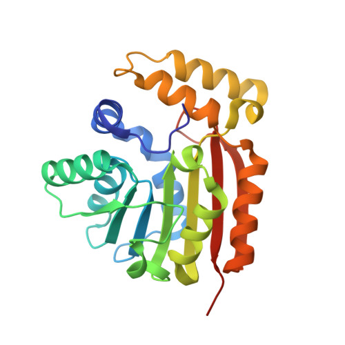
    C-methylation of aromatic small molecules by C-methyltransferases (C-MTs) is an important biological transformation that involves C-C bond formation using S-adenosyl-l-methionine (SAM) as the methyl donor. Here, two advances in the mechanistic understanding of C-methylation of the 8-position of coumarin substrates catalyzed by the C-MT NovO from Streptomyces spheroides are described. First, a crystal structure of NovO reveals the Arg116-Asn117 and His120-Arg121 motifs are essential for coumarin substrate binding. Second, the active-site His120 is responsible for deprotonation of the phenolic 7-hydroxyl group on the coumarin substrate, activating the rate-determining methyl transfer step from SAM. This work expands our mechanistic knowledge of C-MTs, which could be used in the downstream development of engineered biocatalysts for small molecule C-alkylations.


  • Organizational Affiliation

    GlaxoSmithKline Medicines Research Centre , Gunnels Wood Road, Stevenage, SG1 2NY , United Kingdom.


Macromolecules
Find similar proteins by:  (by identity cutoff)  |  3D Structure
Entity ID: 1
MoleculeChains Sequence LengthOrganismDetailsImage
8-demethylnovobiocic acid C(8)-methyltransferase
A, B
236Streptomyces niveusMutation(s): 0 
Gene Names: novO
EC: 2.1.1.284
UniProt
Find proteins for Q9L9F3 (Streptomyces niveus)
Explore Q9L9F3 
Go to UniProtKB:  Q9L9F3
Entity Groups  
Sequence Clusters30% Identity50% Identity70% Identity90% Identity95% Identity100% Identity
UniProt GroupQ9L9F3
Sequence Annotations
Expand
  • Reference Sequence
Experimental Data & Validation

Experimental Data

  • Method: X-RAY DIFFRACTION
  • Resolution: 1.90 Å
  • R-Value Free: 0.202 
  • R-Value Work: 0.178 
  • R-Value Observed: 0.179 
  • Space Group: P 1 21 1
Unit Cell:
Length ( Å )Angle ( ˚ )
a = 43.724α = 90
b = 66.97β = 92.99
c = 80.563γ = 90
Software Package:
Software NamePurpose
REFMACrefinement
XDSdata reduction
Aimlessdata scaling
autoSHARPphasing

Structure Validation

View Full Validation Report


Ligand Structure Quality Assessment 


Entry History 

Deposition Data

Revision History  (Full details and data files)

  • Version 1.0: 2017-01-18
    Type: Initial release
  • Version 1.1: 2017-03-01
    Changes: Database references