5M8E

Crystal structure of a GH43 arabonofuranosidase from Weissella sp. strain 142


Experimental Data Snapshot

  • Method: X-RAY DIFFRACTION
  • Resolution: 2.00 Å
  • R-Value Free: 0.216 
  • R-Value Work: 0.172 
  • R-Value Observed: 0.175 

wwPDB Validation   3D Report Full Report


This is version 1.4 of the entry. See complete history


Literature

Three-dimensional structures and functional studies of two GH43 arabinofuranosidases from Weissella sp. strain 142 and Lactobacillus brevis.

Linares-Pasten, J.A.Falck, P.Albasri, K.Kjellstrom, S.Adlercreutz, P.Logan, D.T.Karlsson, E.N.

(2017) FEBS J 284: 2019-2036

  • DOI: https://doi.org/10.1111/febs.14101
  • Primary Citation of Related Structures:  
    5M8E

  • PubMed Abstract: 

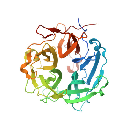
    Arabinofuranosidases degrade arabinose-containing oligo and polysaccharides, releasing l-arabinose, which is a potentially useful sugar, shown to reduce glycemic response under certain conditions. Arabinofuranosidases (Arafs) are frequently found in GH43, one of the most common GH-families encoded in genomes in gut microbiota, and hence it is of interest to increase understanding of the function of these enzymes in species occurring in the gut. Here we have produced, characterized and solved the three-dimensional structures, at 1.9 and 2.0 Å resolution respectively, of two homologous GH43 enzymes, classified under subfamily 26, from Lactobacillus brevis DSM1269 (LbAraf43) and Weissella strain 142 (WAraf43), respectively. The enzymes, with 74% sequence identity to each other, are composed of a single catalytic module with a β-propeller structure typical of GH43, and an active-site pocket with three identifiable subsites (-1, +1, and +2). According to size exclusion chromatography, native WAraf43 is a dimer, while LbAraf43 is a tetramer in solution. Both of them show activity with similar catalytic efficiency on 1,5-α-l-arabinooligosaccharides with a degree of polymerization (DP) of 2-3. Activity is restricted to substrates of low DP, and the reason for this is believed to be an extended loop at the entrance to the active site, creating interactions in the +2 subsite.


  • Organizational Affiliation

    Biotechnology, Department of Chemistry, Lund University, Sweden.


Macromolecules
Find similar proteins by:  (by identity cutoff)  |  3D Structure
Entity ID: 1
MoleculeChains Sequence LengthOrganismDetailsImage
Alpha-N-arabinofuranosidase
A, B
344Weissella cibariaMutation(s): 0 
Gene Names: AO080_02990QX99_00224
EC: 3.2.1.55
UniProt
Find proteins for A0A0D1M7L2 (Weissella cibaria)
Explore A0A0D1M7L2 
Go to UniProtKB:  A0A0D1M7L2
Entity Groups  
Sequence Clusters30% Identity50% Identity70% Identity90% Identity95% Identity100% Identity
UniProt GroupA0A0D1M7L2
Sequence Annotations
Expand
  • Reference Sequence
Experimental Data & Validation

Experimental Data

  • Method: X-RAY DIFFRACTION
  • Resolution: 2.00 Å
  • R-Value Free: 0.216 
  • R-Value Work: 0.172 
  • R-Value Observed: 0.175 
  • Space Group: P 1 21 1
Unit Cell:
Length ( Å )Angle ( ˚ )
a = 59.88α = 90
b = 71.93β = 101.93
c = 79.51γ = 90
Software Package:
Software NamePurpose
BUSTERrefinement
XDSdata reduction
SCALAdata scaling
PHASERphasing

Structure Validation

View Full Validation Report


Entry History & Funding Information

Deposition Data


Funding OrganizationLocationGrant Number
Swedish Research CouncilSweden--

Revision History  (Full details and data files)

  • Version 1.0: 2017-05-10
    Type: Initial release
  • Version 1.1: 2017-05-17
    Changes: Database references
  • Version 1.2: 2017-07-12
    Changes: Data collection, Database references
  • Version 1.3: 2017-09-13
    Changes: Author supporting evidence
  • Version 1.4: 2024-01-17
    Changes: Data collection, Database references, Derived calculations, Refinement description