PELDOR Spectroscopy Reveals Two Defined States of a Sialic Acid TRAP Transporter SBP in Solution.
Glaenzer, J., Peter, M.F., Thomas, G.H., Hagelueken, G.(2017) Biophys J 112: 109-120
- PubMed: 28076802
- DOI: https://doi.org/10.1016/j.bpj.2016.12.010
- Primary Citation of Related Structures:
5LTC - PubMed Abstract:
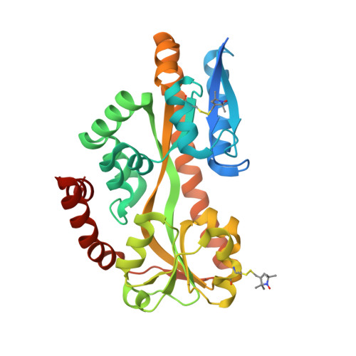
The tripartite ATP-independent periplasmic (TRAP) transporters are a widespread class of membrane transporters in bacteria and archaea. Typical substrates for TRAP transporters are organic acids including the sialic acid N-acetylneuraminic acid. The substrate binding proteins (SBP) of TRAP transporters are the best studied component and are responsible for initial high-affinity substrate binding. To better understand the dynamics of the ligand binding process, pulsed electron-electron double resonance (PELDOR, also known as DEER) spectroscopy was applied to study the conformational changes in the N-acetylneuraminic acid-specific SBP VcSiaP. The protein is the SBP of VcSiaPQM, a sialic acid TRAP transporter from Vibrio cholerae. Spin-labeled double-cysteine mutants of VcSiaP were analyzed in the substrate-bound and -free state and the measured distances were compared to available crystal structures. The data were compatible with two clear states only, which are consistent with the open and closed forms seen in TRAP SBP crystal structures. Substrate titration experiments demonstrated the transition of the population from one state to the other with no other observed forms. Mutants of key residues involved in ligand binding and/or proposed to be involved in domain closure were produced and the corresponding PELDOR experiments reveal important insights into the open-closed transition. The results are in excellent agreement with previous in vivo sialylation experiments. The structure of the spin-labeled Q54R1/L173R1 R125A mutant was solved at 2.1 Å resolution, revealing no significant changes in the protein structure. Thus, the loss of domain closure appears to be solely due to loss of binding. In conclusion, these data are consistent with TRAP SBPs undergoing a simple two-state transition from an open-unliganded to closed-liganded state during the transport cycle.
Organizational Affiliation:
Institute for Physical & Theoretical Chemistry, University of Bonn, Bonn, Germany.