5LCL

STRUCTURE OF the RAD14 DNA-binding domain IN COMPLEX WITH C8-aminofluorene- GUANINE CONTAINING DNA


Experimental Data Snapshot

  • Method: X-RAY DIFFRACTION
  • Resolution: 2.20 Å
  • R-Value Free: 0.256 
  • R-Value Work: 0.217 
  • R-Value Observed: 0.218 

wwPDB Validation   3D Report Full Report


This is version 1.3 of the entry. See complete history


Literature

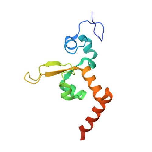
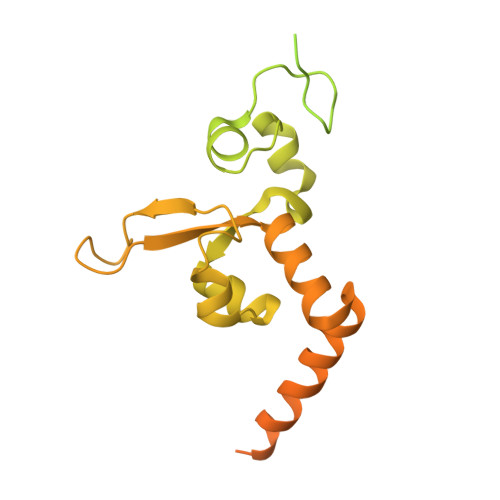
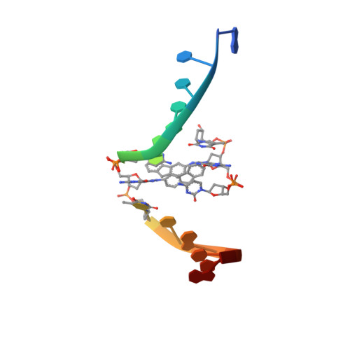
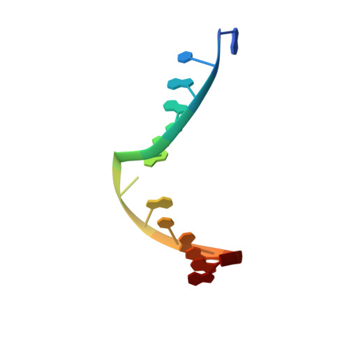
Structural Insights into the Recognition of N(2) -Aryl- and C8-Aryl DNA Lesions by the Repair Protein XPA/Rad14.

Ebert, C.Simon, N.Schneider, S.Carell, T.

(2017) Chembiochem 18: 1379-1382

  • DOI: https://doi.org/10.1002/cbic.201700169
  • Primary Citation of Related Structures:  
    5LCL, 5LCM

  • PubMed Abstract: 

    Aromatic amines are strongly carcinogenic. They are activated in the liver to give reactive nitrenium ions that react with nucleobases within the DNA duplex. The reaction occurs predominantly at the C8 position of the dG base, thereby giving C8-acetyl-aryl- or C8-aryl-dG adducts in an electrophilic aromatic substitution reaction. Alternatively, reaction with the exocyclic 2-NH 2 group is observed. Although the C8 adducts retain base-pairing properties, base pairing is strongly compromised in the case of the N 2 adducts. Here we show crystal structures of two DNA lesions, N 2 -acetylnaphthyl-dG and C8-fluorenyl-dG, within a DNA duplex recognized by the repair protein Rad14. The structures confirm that two molecules of the repair protein recognize the lesion and induce a 72 or 78° kink at the site of the damage. Importantly, the same overall kinked structure is induced by binding of the repair proteins, although the structurally different lesions result in distinct stacking interactions of the lesions within the duplex. The results suggest that the repair protein XPA/Rad14 is a sensor that recognizes flexibility. The protein converts the information that structurally different lesions are present in the duplex into a unifying sharply kinked recognition motif.


  • Organizational Affiliation

    Center for Integrated Protein Science at the Department of Chemistry, Ludwig-Maximilians-Universität München, Butenandtstrasse 5-13, 81377, München, Germany.


Macromolecules

Find similar proteins by:  (by identity cutoff)  |  3D Structure
Entity ID: 1
MoleculeChains Sequence LengthOrganismDetailsImage
DNA repair protein RAD14119Saccharomyces cerevisiae S288CMutation(s): 0 
Gene Names: RAD14YMR201CYM8325.02C
UniProt
Find proteins for P28519 (Saccharomyces cerevisiae (strain ATCC 204508 / S288c))
Explore P28519 
Go to UniProtKB:  P28519
Entity Groups  
Sequence Clusters30% Identity50% Identity70% Identity90% Identity95% Identity100% Identity
UniProt GroupP28519
Sequence Annotations
Expand
  • Reference Sequence
Find similar proteins by:  (by identity cutoff)  |  3D Structure
Entity ID: 2
MoleculeChains Sequence LengthOrganismDetailsImage
DNA repair protein RAD14371Saccharomyces cerevisiae S288CMutation(s): 0 
Gene Names: RAD14YMR201CYM8325.02C
UniProt
Find proteins for P28519 (Saccharomyces cerevisiae (strain ATCC 204508 / S288c))
Explore P28519 
Go to UniProtKB:  P28519
Entity Groups  
Sequence Clusters30% Identity50% Identity70% Identity90% Identity95% Identity100% Identity
UniProt GroupP28519
Sequence Annotations
Expand
  • Reference Sequence

Find similar nucleic acids by:  Sequence   |   3D Structure  

Entity ID: 3
MoleculeChains LengthOrganismImage
GCTCTAC(8AF)TCATCA15Saccharomyces
Sequence Annotations
Expand
  • Reference Sequence

Find similar nucleic acids by:  Sequence   |   3D Structure  

Entity ID: 4
MoleculeChains LengthOrganismImage
DNA (5'-D(*GP*TP*GP*AP*TP*GP*AP*CP*GP*TP*AP*GP*AP*G)-3')15Saccharomyces
Sequence Annotations
Expand
  • Reference Sequence
Experimental Data & Validation

Experimental Data

  • Method: X-RAY DIFFRACTION
  • Resolution: 2.20 Å
  • R-Value Free: 0.256 
  • R-Value Work: 0.217 
  • R-Value Observed: 0.218 
  • Space Group: P 41
Unit Cell:
Length ( Å )Angle ( ˚ )
a = 53.205α = 90
b = 53.205β = 90
c = 130.965γ = 90
Software Package:
Software NamePurpose
REFMACrefinement
XDSdata reduction
XDSdata scaling
REFMACphasing

Structure Validation

View Full Validation Report


Entry History & Funding Information

Deposition Data


Funding OrganizationLocationGrant Number
German Research FoundationGermany--

Revision History  (Full details and data files)

  • Version 1.0: 2017-05-10
    Type: Initial release
  • Version 1.1: 2017-06-07
    Changes: Database references
  • Version 1.2: 2017-07-26
    Changes: Database references
  • Version 1.3: 2024-01-10
    Changes: Data collection, Database references, Derived calculations, Refinement description