


Novel Biochemical and Structural Insights into the Interaction of Myristoylated Cargo with Unc119 Protein and Their Release by Arl2/3.
Jaiswal, M., Fansa, E.K., Kosling, S.K., Mejuch, T., Waldmann, H., Wittinghofer, A.(2016) J Biol Chem 291: 20766-20778
- PubMed: 27481943
- DOI: https://doi.org/10.1074/jbc.M116.741827
- Primary Citation of Related Structures:
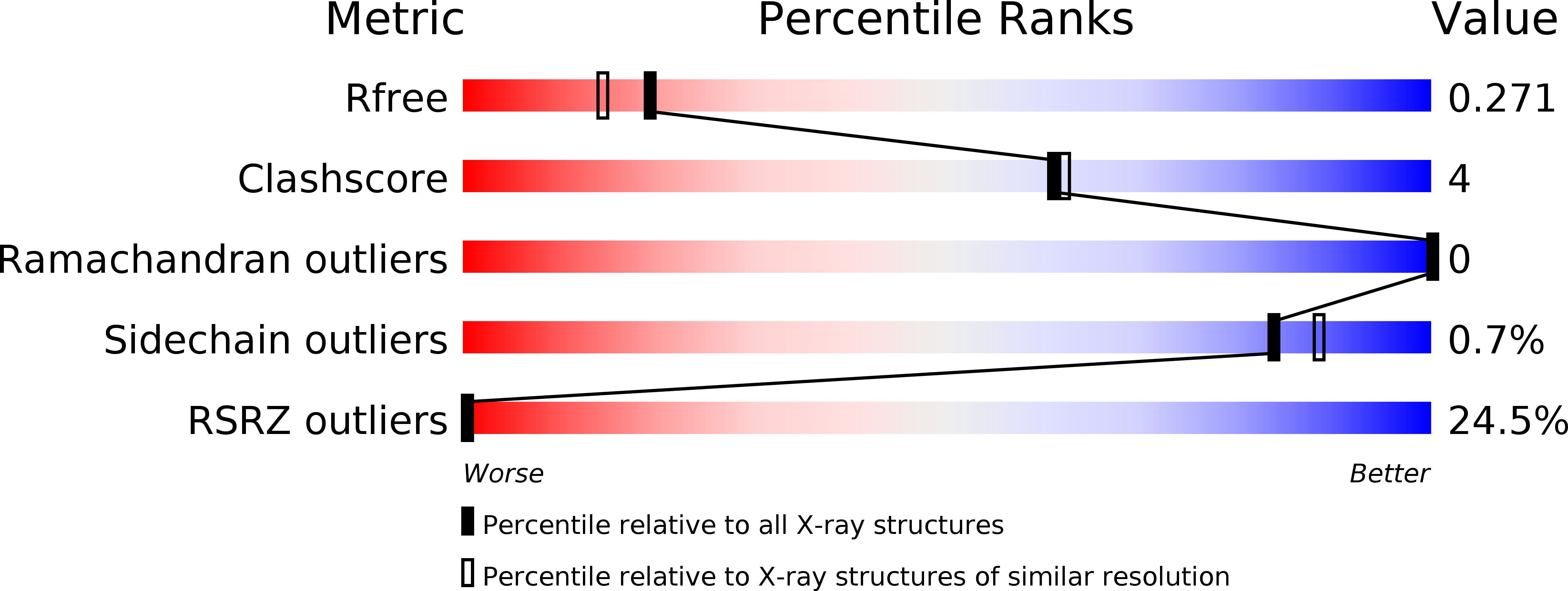
5L7K - PubMed Abstract:
Primary cilia are highly specialized small antenna-like cellular protrusions that extend from the cell surface of many eukaryotic cell types. The protein content inside cilia and cytoplasm is very different, but details of the sorting process are not understood for most ciliary proteins. Recently, we have shown that prenylated proteins are sorted according to their affinity to the carrier protein PDE6δ and the ability of Arl3 but not Arl2 to release high affinity cargo inside the cilia (Fansa, E. K., Kösling, S. K., Zent, E., Wittinghofer, A., and Ismail, S. (2016) Nat. Commun. 7, 11366). Here we address the question whether a similar principle governs the transport of myristoylated cargo by the carrier proteins Unc119a and Unc119b. We thus analyzed the binding strength of N-terminal myristoylated cargo peptides (GNAT1, NPHP3, Cystin1, RP2, and Src) to Unc119a and Unc119b proteins. The affinity between myristoylated cargo and carrier protein, Unc119, varies between subnanomolar and micromolar. Peptides derived from ciliary localizing proteins (GNAT1, NPHP3, and Cystin1) bind with high affinity to Unc119 proteins, whereas a peptide derived from a non-ciliary localizing protein (Src) has low affinity. The peptide with intermediate affinity (RP2) is localized at the ciliary transition zone as a gate keeper. We show that the low affinity peptides are released by both Arl2·GppNHp and Arl3·GppNHp, whereas the high affinity peptides are exclusively released by only Arl3·GppNHp. Determination of the x-ray structure of myristoylated NPHP3 peptide in complex with Unc119a reveals the molecular details of high affinity binding and suggests the importance of the residues at the +2 and +3 positions relative to the myristoylated glycine for high and low affinities. The mutational analysis of swapping the residues at the +2 and +3 positions between high and low affinity peptides results in reversing their affinities for Unc119a and leads to a partial mislocalization of a low affinity mutant of NPHP3.
Organizational Affiliation:
From the Structural Biology Group and.


