


Crystal Structure of Human Pseudouridylate Synthase 7
ZENG, H., DONG, A., WALKER, J.R., Bountra, C., Arrowsmith, C.H., Edwards, A.M., BROWN, P.J., WU, H., Structural Genomics Consortium (SGC)To be published.
Experimental Data Snapshot
wwPDB Validation 3D Report Full Report

Entity ID: 1 | |||||
|---|---|---|---|---|---|
| Molecule | Chains | Sequence Length | Organism | Details | Image |
| Pseudouridylate synthase 7 | 563 | Homo sapiens | Mutation(s): 0 Gene Names: PUS7, KIAA1897 EC: 5.4.99 | 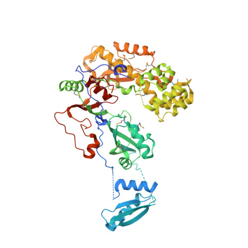 | |
UniProt & NIH Common Fund Data Resources | |||||
Find proteins for Q96PZ0 (Homo sapiens) Explore Q96PZ0 Go to UniProtKB: Q96PZ0 | |||||
PHAROS: Q96PZ0 GTEx: ENSG00000091127 | |||||
Entity Groups | |||||
| Sequence Clusters | 30% Identity50% Identity70% Identity90% Identity95% Identity100% Identity | ||||
| UniProt Group | Q96PZ0 | ||||
Sequence AnnotationsExpand | |||||
| |||||
| Ligands 2 Unique | |||||
|---|---|---|---|---|---|
| ID | Chains | Name / Formula / InChI Key | 2D Diagram | 3D Interactions | |
| EDO Query on EDO | B [auth A] | 1,2-ETHANEDIOL C2 H6 O2 LYCAIKOWRPUZTN-UHFFFAOYSA-N |  | ||
| UNX Query on UNX | C [auth A], D [auth A], E [auth A] | UNKNOWN ATOM OR ION X |  | ||
| Modified Residues 1 Unique | |||||
|---|---|---|---|---|---|
| ID | Chains | Type | Formula | 2D Diagram | Parent |
| MSE Query on MSE | A | L-PEPTIDE LINKING | C5 H11 N O2 Se |  | MET |
| Length ( Å ) | Angle ( ˚ ) |
|---|---|
| a = 56.356 | α = 90 |
| b = 73.4 | β = 90 |
| c = 138.179 | γ = 90 |
| Software Name | Purpose |
|---|---|
| BUSTER-TNT | refinement |
| SCALEPACK | data scaling |
| PDB_EXTRACT | data extraction |
| Coot | model building |
| HKL-3000 | data reduction |
| RESOLVE | phasing |
| WARP | model building |
RCSB PDB (citation) is hosted by
RCSB PDB is a member of the

