Structural and In Vivo Studies on Trehalose-6-Phosphate Synthase from Pathogenic Fungi Provide Insights into Its Catalytic Mechanism, Biological Necessity, and Potential for Novel Antifungal Drug Design.
Miao, Y., Tenor, J.L., Toffaletti, D.L., Maskarinec, S.A., Liu, J., Lee, R.E., Perfect, J.R., Brennan, R.G.(2017) mBio 8
- PubMed: 28743811
- DOI: https://doi.org/10.1128/mBio.00643-17
- Primary Citation of Related Structures:
5HUT, 5HUU, 5HVL, 5HVM, 5HVO - PubMed Abstract:
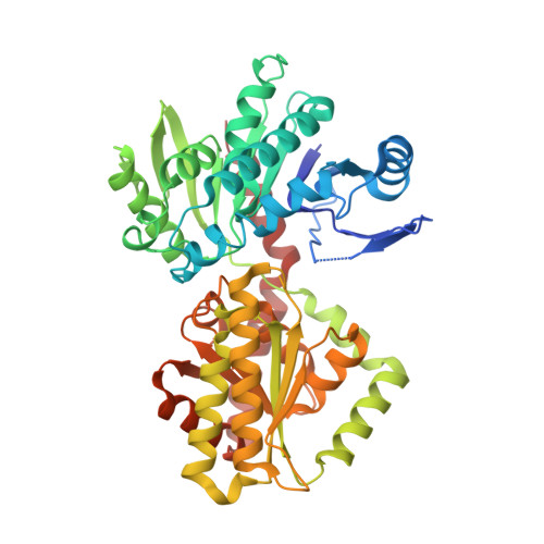
The disaccharide trehalose is critical to the survival of pathogenic fungi in their human host. Trehalose-6-phosphate synthase (Tps1) catalyzes the first step of trehalose biosynthesis in fungi. Here, we report the first structures of eukaryotic Tps1s in complex with substrates or substrate analogues. The overall structures of Tps1 from Candida albicans and Aspergillus fumigatus are essentially identical and reveal N- and C-terminal Rossmann fold domains that form the glucose-6-phosphate and UDP-glucose substrate binding sites, respectively. These Tps1 structures with substrates or substrate analogues reveal key residues involved in recognition and catalysis. Disruption of these key residues severely impaired Tps1 enzymatic activity. Subsequent cellular analyses also highlight the enzymatic function of Tps1 in thermotolerance, yeast-hypha transition, and biofilm development. These results suggest that Tps1 enzymatic functionality is essential for the fungal stress response and virulence. Furthermore, structures of Tps1 in complex with the nonhydrolyzable inhibitor, validoxylamine A, visualize the transition state and support an internal return-like catalytic mechanism that is generalizable to other GT-B-fold retaining glycosyltransferases. Collectively, our results depict key Tps1-substrate interactions, unveil the enzymatic mechanism of these fungal proteins, and pave the way for high-throughput inhibitor screening buttressed and guided by the current structures and those of high-affinity ligand-Tps1 complexes. IMPORTANCE Invasive fungal diseases have emerged as major threats, resulting in more than 1.5 million deaths annually worldwide. This epidemic has been further complicated by increasing resistance to all major classes of antifungal drugs in the clinic. Trehalose biosynthesis is essential for the fungal stress response and virulence. Critically, this biosynthetic pathway is absent in mammals, and thus, the two enzymes that carry out trehalose biosynthesis, namely, trehalose-6-phosphate synthase (Tps1) and trehalose-6-phosphate phosphatase (Tps2), are prominent targets for antifungal intervention. Here, we report the first eukaryotic Tps1 structures from the pathogenic fungi Candida albicans and Aspergillus fumigatus in complex with substrates, substrate analogues, and inhibitors. These structures reveal key protein-substrate interactions, providing atomic-level scaffolds for structure-guided drug design of novel antifungals that target Tps1.
Organizational Affiliation:
Department of Biochemistry, Duke University School of Medicine, Durham, North Carolina, USA.